

Image-guided oncologic surgery using invisible light: completed pre-clinical development for sentinel lymph node mapping
- PMID: 17009138
- PMCID: PMC2474791
- DOI: 10.1245/s10434-006-9194-6
Image-guided oncologic surgery using invisible light: completed pre-clinical development for sentinel lymph node mapping
Abstract
Background: Invisible near-infrared (NIR) fluorescent light permits high sensitivity, real-time image-guidance during oncologic surgery without changing the look of the surgical field. In this study, we complete pre-clinical development of the technology for sentinel lymph node (SLN) mapping using a large animal model of spontaneous melanoma.
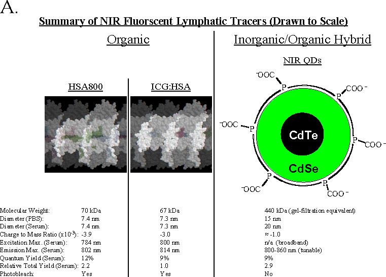
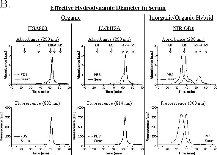
Methods: Sinclair swine with spontaneous melanoma metastatic to regional lymph nodes were used because of their similarity to human melanoma. Organic lymphatic tracers tested included FDA-approved indocyanine green adsorbed non-covalently to human serum albumin (HSA), and NIR fluorophore CW800 conjugated covalently to HSA (HSA800). The inorganic/organic hybrid tracer tested was type II NIR quantum dots with an anionic coating. Primary tumors received four peri-tumoral injections of each tracer, with a fluorophore dose of 100 pmol to 1 nmol per injection. SLN mapping and image-guided resection were performed in real-time.
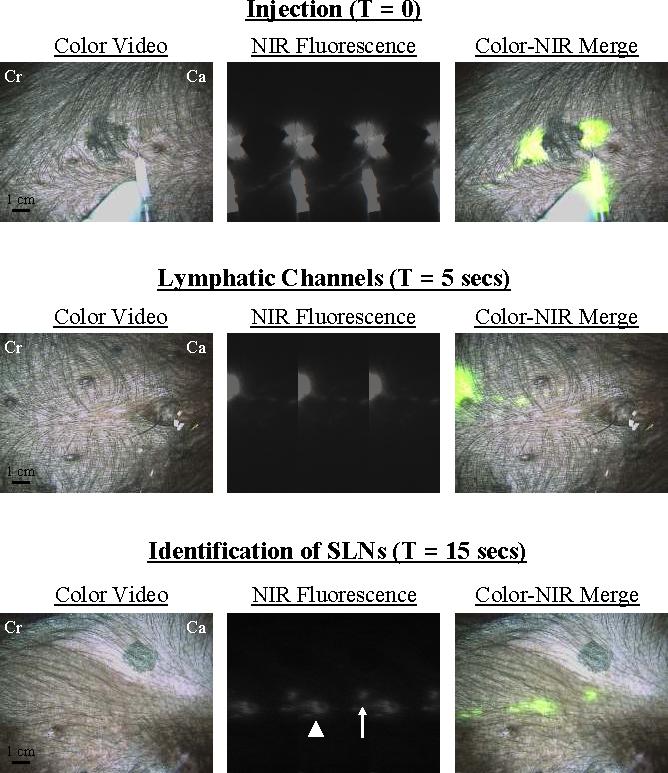
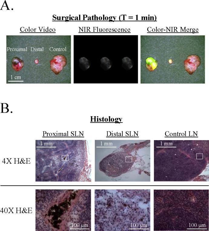
Results: Each of the 3 lymphatic tracers was injected into n = 4 separate primary melanomas in a total of 6 animals. All 12 injections resulted in identification of the SLN(s) and their associated lymphatic channels within 1 minute in 100% of cases, despite highly pigmented skin and black fur. Hydrodynamic diameter had a profound impact on tracer behavior in vivo.
Conclusions: This study completes the pre-clinical development of NIR fluorescence-guided SLN mapping and provides insight into imaging system optimization and tracer choice for future human clinical trials. The technology is likely to eliminate the need for radioactive and colored tracers, permits real-time image guidance throughout the procedure, and assists the pathologist in tissue analysis.
Figures
References
-
- Lim YT, Kim S, Nakayama A, Stott NE, Bawendi MG, Frangioni JV. Selection of quantum dot wavelengths for biomedical assays and imaging. Mol Imaging. 2003;2:50–64. - PubMed
-
- Frangioni JV. In vivo near-infrared fluorescence imaging. Curr Opin Chem Biol. 2003;7:626–34. - PubMed
-
- Zaheer A, Lenkinski RE, Mahmood A, Jones AG, Cantley LC, Frangioni JV. In vivo near-infrared fluorescence imaging of osteoblastic activity. Nat Biotechnol. 2001;19:1148–54. - PubMed
-
- Nakayama A, del Monte F, Hajjar RJ, Frangioni JV. Functional near-infrared fluorescence imaging for cardiac surgery and targeted gene therapy. Mol Imaging. 2002;1:365–77. - PubMed
-
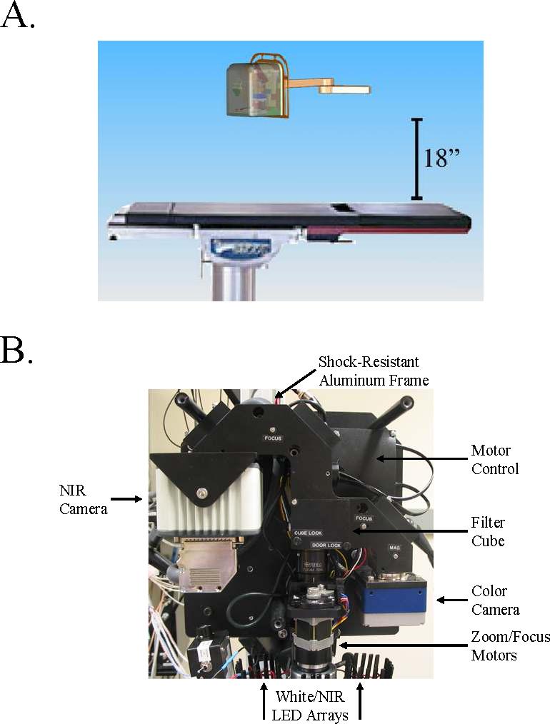
- De Grand AM, Frangioni JV. An operational near-infrared fluorescence imaging system prototype for large animal surgery. Technol Cancer Res Treat. 2003;2:553–62. - PubMed
Publication types
MeSH terms
Substances
Grants and funding
LinkOut - more resources
Full Text Sources
Other Literature Sources
Medical
Miscellaneous
