Comparative Study
. 2006 Autumn;11(3):216-26.
doi: 10.1379/csc-191r.1.

Fusion proteins of Hsp70 with tumor-associated antigen acting as a potent tumor vaccine and the C-terminal peptide-binding domain of Hsp70 being essential in inducing antigen-independent anti-tumor response in vivo

Affiliations
Comparative Study

Fusion proteins of Hsp70 with tumor-associated antigen acting as a potent tumor vaccine and the C-terminal peptide-binding domain of Hsp70 being essential in inducing antigen-independent anti-tumor response in vivo

Honghai Zhang et al. Cell Stress Chaperones. 2006 Autumn.

Abstract

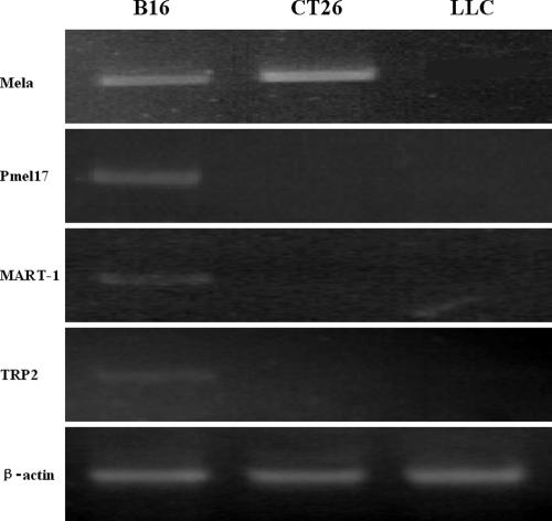
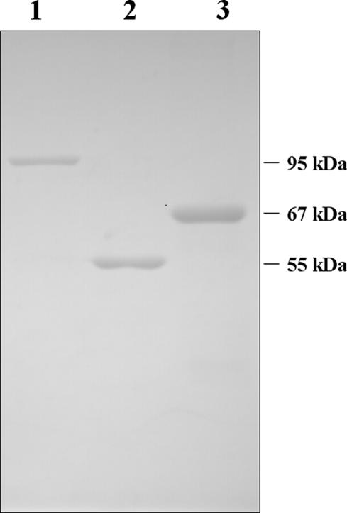
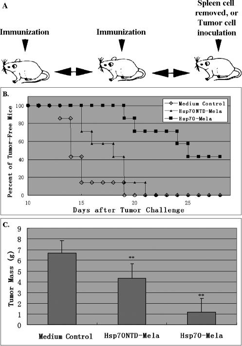
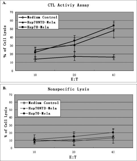
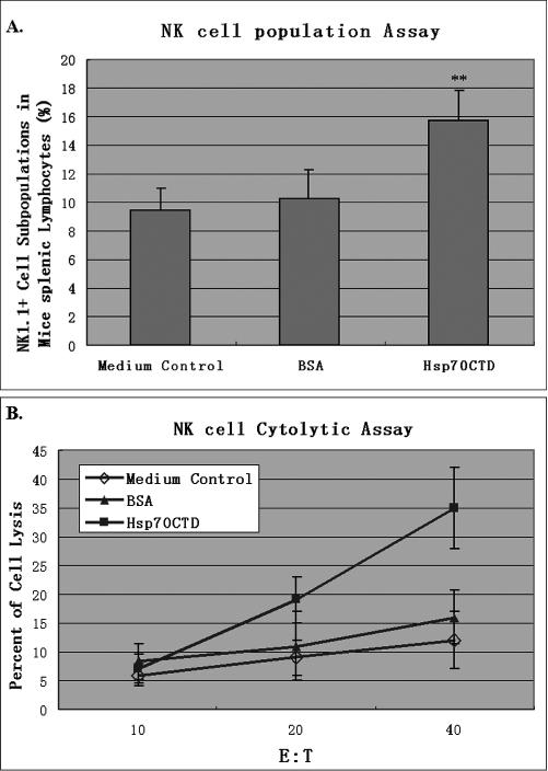
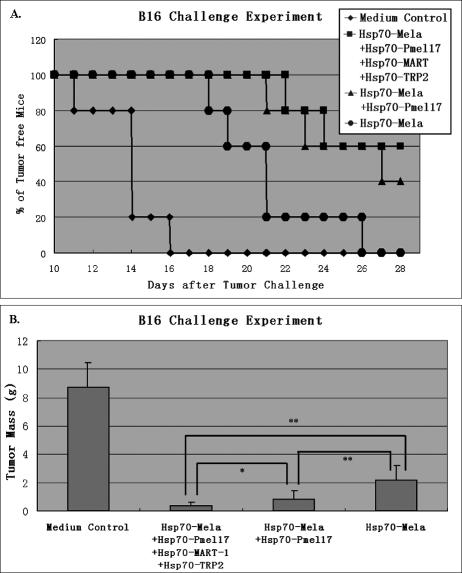
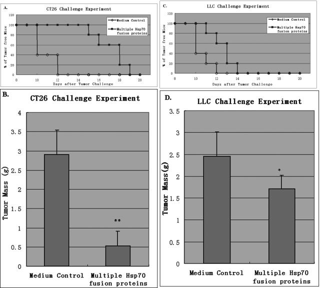
Hsp70s are a family of ATP-dependent chaperones of relative molecular mass around 70 kDa. Immunization of mice with Hsp70 isolated from tumor tissues has been proved to elicit specific protective immunity against the original tumor challenge. In this work, we investigated whether Hsp70 can be used as vehicle to elicit immune response to its covalence-accompanying antigen. A recombinant protein expression vector was constructed that permitted the production of recombinant protein fusing tumor-associated antigen (eg, Mela) to the C terminus of Hsp70. We found that the Hsp70-Mela fusion protein can elicit strong cellular immune responses against murine tumor B16, which expresses protein Mela. The Hsp70 peptide-binding domain deletion mutant of the fusion protein was sufficient for inducing Mela-specific cytotoxic T lymphocyte but was not sufficient for engendering potent anti-tumor immunity against B16. We also found that host natural killer (NK) cells were stimulated in vivo by C-terminal domain of Hsp70. We thus presume that Hsp70 fusion proteins suppress tumor growth via at least 2 distinct pathways: one is covalence-accompanying antigen dependent; another is antigen independent. The C-terminal domain of Hsp70 seemed to be the crucial part in eliciting antigen-independent responses, including NK cell stimulation, against tumor challenges. Furthermore, we found that immunization with multiple Hsp70 fusion proteins resulted in a better anti-tumor effect.


Figures

Fig 1.
Fig 1.
Antigen expression analysis. The mRNAs of Mela, Pmel17, TRP2, and MART-1 were detected in B16 cells by RT-PCR; none of the 4 antigen mRNAs were detected in LLC cells, and only Mela mRNA was detected in CT26 cells
Fig 2.
Fig 2.
Production and purification of recombinant proteins. Purified proteins were examined by SDS-PAGE and visualized by Coomassie staining. Lane 1, Hsp70–Mela; lane 2, GST– Hsp70CTD; lane 3, Hsp70NTD–Mela. Molecular mass markers are on the right side
Fig 3.
Fig 3.
Suppressive effects against B16. Groups of mice (n = 7 per group) were immunized with Hsp70–Mela (filled squares), Hsp70NTD–Mela (filled triangles), or medium control (open diamonds) twice before tumor challenge by the injection schedule (A). Kaplan–Meier curves (B) showed that the tumor-free survival interval was 24.9 ± 1.2 days in the Hsp70–Mela immunized group, 17.6 ± 0.9 in the Hsp70NTD–Mela immunized group, and 14.9 ± 0.7 days in the control group. Comparison of animal survival curves was performed by log-rank test. Hsp70–Mela versus control (P = 0.0002), Hsp70NTD–Mela versus control (P = 0.043), Hsp70-Mela versus Hsp70NTD–Mela (P = 0.001). (C) Tumor tissue weights of the 3 groups of mice on day 28 after tumor challenge. Each mean tumor weight of a mouse injected with Hsp70N–Mela and Hsp70– Mela decreased significantly compared with the control group (P < 0.001). Results are expressed as the mean ± standard deviation (error bars). ** P < 0.01 versus the control group. Data represent 3 experiments
Fig 4.
Fig 4.
Cytotoxicity T-lymphocyte (CTL) assay. Groups of mice (n = 3) were immunized twice with Hsp70–Mela (filled squares), Hsp70NTD–Mela (filled triangles), or medium control (open diamonds). Mouse splenic lymphocytes were collected 1 week after the boost then stimulated for 5 days with mitomycin C–treated B16 cells. Cytotoxicity was measured by LDH release assay. B16 cells were used as target cells. Stimulated splenocytes were incubated with B16 cells at the indicated effector;th:;thtarget cell ratio (E/T) for 4 hours at 37°C. Similar specific lysis was observed in Hsp70–Mela and Hsp70NTD–Mela immunization groups, either of which is statistically significant compared with the control (A). The nonspecific lysis assay was also performed, in which the Mela-negative LLC acted as target cell. No significant nonspecific lysis was observed (B). Results are expressed as the mean ± standard deviation (error bars)
Fig 5.
Fig 5.
NK cell stimulatory assay. Groups of C57BL/6 mice (n = 5 per group) were injected with 0.15 nmol of Hsp70CTD (filled squares), BSA (filled triangles), or PBS (open diamonds) subcutaneously twice at 1-week interval. Splenic lymphocytes were collected 1 week after the second injection and subsequently stained with PE-conjugated anti-mouse NK1.1 antibody; the NK cell populations were determined via flow cytometry (A). There is an increased splenic NK cell population in the mice immunized with Hsp70CTD (P < 0.01). NK cell cytotoxicities of the splenic lymphocytes were also determined (B) via LDH release assay, in which YAC-1 cells acted as target cells. The results indicated that splenic lymphocytes from the group of mice injected with Hsp70CTD but not with BSA had significantly increased NK cell cytotoxicities against YAC-1 (P < 0.01). Results are expressed as the mean ± standard deviation (error bars). ** P < 0.01 compared with the control group
Fig 6.
Fig 6.
Suppressive effect of multiple Hsp70 fusion proteins against B16. In the B16 challenge experiment, groups of C57BL/6 mice (n = 5 per group) were immunized with Hsp70–Mela only (filled cycles), a mixture of Hsp70–Mela and Hsp70–Pmel17 (filled triangles), or a mixture of 4 Hsp70 fusion proteins (Hsp70–Mela, Hsp70– Pmel17, Hsp70–MART-1, and Hsp70–TRP2; filled squares) twice before inoculation of 2 × 10⁶ B16 melanoma cells. Each dose of the 4 fusion proteins is 0.15 nmol, and a medium control group (filled diamonds) was set. Kaplan–Meier curves (A) showed that the tumor-free survival interval was 26.0 ± 1.1 days in the 4 Hsp70 fusion protein immunization group, 25.4 ± 1.3 in the 2 Hsp70 fusion protein immunization group, 21.0 ± 1.4 in the Hsp70–Mela immunization group, and 13.8 ± 0.8 days in the control group. Comparison of animal survival curves was performed by log-rank test. The Hsp70– Mela group versus the 2 Hsp70 fusion proteins group (P = 0.033), Hsp70–Mela group versus the 4 Hsp70 fusion proteins group (P = 0.020), and the 2 Hsp70 fusion proteins group versus the 4 Hsp70 fusion proteins group (P = 0.57). At day 28 after tumor challenge, B16 melanoma nodules were excised away from the challenged mice and weighed. (B) Each of the 3 groups immunized with Hsp70 fusion proteins had a lower mean tumor weight than control group (P < 0.01). Among the 3 groups, the mean tumor weight of the group immunized with 4 Hsp70 fusion proteins at one time was significantly less than the group immunized with Hsp70–Mela only (P < 0.01) and the group immunized with Hsp70–Mela and Hsp70–Pmel17 (0.01 < P < 0.05), which was lighter than the group immunized with Hsp70–Mela only (P < 0.01). Results are expressed as the mean ± standard deviation (error bars). * P < 0.01, ** 0.01 < P < 0.05. Data are representative of 2 experiments
Fig 7.
Fig 7.
Suppressive effect of multiple Hsp70 fusion proteins against CT26 and LLC. Groups of BABL/c (or C57BL/6) mice (n = 5 per group) were immunized with the mixture of multiple Hsp70 fusion proteins (filled squares) or PBS (opened squares) twice before inoculation of 1 × 10⁴ CT26 (or 5 × 10⁴ LLC) cells. (A) Kaplan–Meier curves were generated to show the tumor-free survival interval in each group of the CT26 challenge experiment: 17.0 ± 0.6 days in the immunized group and 10.8 ± 0.5 days in the control group. The difference between the 2 groups was significant (P = 0.002). These mice were sacrificed on day 20 after the tumor challenge and tumor nodules were removed and weighed. (B) The CT26 tumor mass was reduced by 87% in the immunized group compared with the control group (P < 0.01). (C) Kaplan– Meier curves were generated to show the tumor-free survival interval of each group in the LLC challenge experiment: 12.6 ± 0.5 days in the immunized group and 10.6 ± 0.4 days in the control group. The difference between the 2 groups was significant (P = 0.002). These mice were sacrificed on day 20 after the tumor challenge, and tumor nodules were removed and weighed. (D) The LLC tumor mass was reduced by 29% in the immunized group compared with the control group (0.01 < P < 0.05). Tumor masses are expressed as the mean ± standard deviation (error bars). * P < 0.01, ** 0.01 < P < 0.05. Data are representative of 3 experiments

Similar articles

Cited by

References

    1. Asea A, Kraeft SK, Kurt-Jones EA, Stevenson MA, Chen LB, Finberg RW, Koo GC, Calderwood SK. Hsp70 stimulates cytokine production through a CD14-dependant pathway, demonstrating its dual role as a chaperone and cytokine. Nat Med. 2000;6:435–442.1078-8956(2000)006[0435:HSCPTA]2.0.CO;2 - PubMed
    1. Belli F, Testori A, and Rivoltini L. et al. 2002 Vaccination of metastatic melanoma patients with autologous tumor-derived heat shock protein gp96-peptide complexes: clinical and immunologic findings. J Clin Oncol. 20:4169–4180. - PubMed
    1. Binder RJ, Vatner R, Srivastava P. The heat-shock protein receptors: some answers and more questions. Tissue Antigens. 2004;64:442–451.0001-2815(2004)064[0442:THPRSA]2.0.CO;2 - PubMed
    1. Blachere NE, Li ZH, Chandawarkar RY, Suto R, Jaikaria NS, Basu S, Udono H, Srivastava PK. Heat shock protein–peptide complexes, reconstituted in vitro, elicit peptide-specific cytotoxic T lymphocyte response and tumor immunity. J Exp Med. 1997;186:1315–1322.0022-1007(1997)186[1315:HSPCRI]2.0.CO;2 - PMC - PubMed
    1. Bloom MB, PerryLalley D, Robbins PF, Li Y, ElGamil M, Rosenberg SA, Yang JC. Identification of tyrosinase-related protein 2 as a tumor rejection antigen for the B16 melanoma. J Exp Med. 1997;185:453–459.0022-1007(1997)185[0453:IOTPAA]2.0.CO;2 - PMC - PubMed

Publication types

MeSH terms

LinkOut - more resources