. 2006 Sep 29;2(9):e134.
doi: 10.1371/journal.pcbi.0020134. Epub 2006 Aug 23.

Targeted molecular dynamics study of C-loop closure and channel gating in nicotinic receptors

Affiliations

Targeted molecular dynamics study of C-loop closure and channel gating in nicotinic receptors

Xiaolin Cheng et al. PLoS Comput Biol. .

Abstract

The initial coupling between ligand binding and channel gating in the human alpha7 nicotinic acetylcholine receptor (nAChR) has been investigated with targeted molecular dynamics (TMD) simulation. During the simulation, eight residues at the tip of the C-loop in two alternating subunits were forced to move toward a ligand-bound conformation as captured in the crystallographic structure of acetylcholine binding protein (AChBP) in complex with carbamoylcholine. Comparison of apo- and ligand-bound AChBP structures shows only minor rearrangements distal from the ligand-binding site. In contrast, comparison of apo and TMD simulation structures of the nAChR reveals significant changes toward the bottom of the ligand-binding domain. These structural rearrangements are subsequently translated to the pore domain, leading to a partly open channel within 4 ns of TMD simulation. Furthermore, we confirmed that two highly conserved residue pairs, one located near the ligand-binding pocket (Lys145 and Tyr188), and the other located toward the bottom of the ligand-binding domain (Arg206 and Glu45), are likely to play important roles in coupling agonist binding to channel gating. Overall, our simulations suggest that gating movements of the alpha7 receptor may involve relatively small structural changes within the ligand-binding domain, implying that the gating transition is energy-efficient and can be easily modulated by agonist binding/unbinding.


Conflict of interest statement

Competing interests. The authors have declared that no competing interests exist.

Figures

Figure 1
Figure 1. Amino Acid Sequence of the Human α7 Nicotinic Receptor Aligned with AChBP and the Torpedo Receptor
Sequence numbering corresponds to the human α7 receptor. Positions colored red and green are highly conserved, and have been implicated in the channel gating mechanism.
Figure 2
Figure 2. Structural Comparison of apo and ACh-Bound Conformations of the Ligand Binding Domain of Human α7 Receptor
The triad of conserved residues is shown in stick representation. The tip of C-loop, to which the targeted force is applied (residues Arg186-Glu193), is colored orange. The apo (A) conformation is the homology model based on the Torpedo α subunit, while the ACh-bound (B) conformation is modeled based on the crystal structure of AChBP with bound carbamoylcholine [8]; (C) the PMF for the interaction between Lys145 and Tyr188 (see text).
Figure 3
Figure 3. Structural Changes of β10, Cys loop, and β1–β2 loop during the TMD Simulation
Comparison of the initial structure (silver) and a snapshot from the last 50 ps of the TMD simulation (green), (A) front view; (B) side view rotated 120° with respect to view (A).
Figure 4
Figure 4. The Hydrogen Bond Distances between Arg206 and Glu45
(A) During the 10-ns control simulation. (B) During the 4-ns TMD simulation. Detailed view of residues Arg206, Glu45, Lys46, and Pro269 in the starting structure (C) and in the final conformation from the TMD simulation (D).
Figure 5
Figure 5. Energetic and Structural Changes of Several Key Residues at the Membrane Interface during the TMD Simulation
(A) Estimation of the nonbonded interaction energies between Glu45, Lys46, and Pro269 during the TMD simulation. (B) Cα(R206) –Cα(E45) distance as a function of time during the TMD simulation.
Figure 6
Figure 6. The RMS Fluctuations of Cα Atoms during the TMD Simulation
For clarity, only results for subunit A are shown. The inset shows the displacements of the β9 and β10 strands. Asp197 remains relatively stationary during the enforced inward motion of the C-loop, as indicated by its low displacement value.
Figure 7
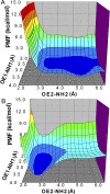
Figure 7. The PMFs for the Interaction between Glu45 and Arg206
(A) In the starting structure. (B) In the final conformer from the TMD simulation. PMF energy is shown on a continuous color scale from 0 (blue) to ≥ 10 kcal/mol (red).
Figure 8
Figure 8. Minimum Pore Radius and z-Axis Position of the Minimum Pore Radius as a Function of Time
(A,C) During the control simulation. (B,D) During the TMD simulation. The center of the pore domain is set to zero, with positive z values toward the extracellular end. The conventional numbering for M2 residues is depicted on the right axis.
Figure 9
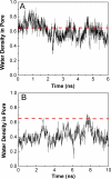
Figure 9. Water Density in the Central 5-Å Portion of the Pore Normalized by the Bulk Water Density
(A) During the simulations of the final TMD conformer. (B) During the simulations of the control simulation. The red dashed lines indicate the water density at 0.65 of the bulk density.
Figure 10
Figure 10. The Iso-Surfaces of Time-Averaged Water Density
(A) During the control simulation. (B) During the the simulation commencing from the final TMD conformer. The surface corresponds to the iso-density contour ~0.4 of the bulk water density. Five M2 helices are shown in ribbons. Residue Val252 is highlighted as yellow spheres and displaced sideways in the final TMD conformer, contributing to the higher water density observed for this simulation.
Figure 11
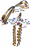
Figure 11. Sequence of Conformational Changes in the Ligand-Binding and Transmembrane Domains of the Human α7 Receptor
The resting and activated conformations are shown in silver and orange, respectively. Glu45, Lys46, Lys145, Tyr188, and Arg206 are shown in ball-and-stick representation. Pro269 is shown in sphere representation.
Figure 12
Figure 12. A Schematic Representation of Helix Tilting, Rotation around the Channel Axis, and Self-Rotation Angles
(A) Helix tilting angle. (B) Rotation around the channel axis angle. (C) Self-rotation angle.

References

    1. Alberts B, Johnson A, Lewis J, Raff M, Roberts K, et al. Molecular biology of the cell. 4th edition. New York: Garland Publishing; 2002. 1616
    1. Karlin A. Emerging structure of the nicotinic acetylcholine receptors. Nat Rev Neurosci. 2002;3:102–114. - PubMed
    1. Jensen AA, Frolund B, Liljefors T, Krogsgaard-Larsen P. Neuronal nicotinic acetylcholine receptors: Structural revelations, target identifications, and therapeutic inspirations. J Med Chem. 2005;48:4705–4745. - PubMed
    1. Cassels BK, Bermudez I, Dajas F, Abin-Carriquiry JA, Wonnacott S. From ligand design to therapeutic efficacy: The challenge for nicotinic receptor research. Drug Discov Today. 2005;10:1657–1665. - PubMed
    1. Dajas-Bailador F, Wonnacott S. Nicotinic acetylcholine receptors and the regulation of neuronal signalling. Trends Pharmacol Sci. 2004;25:317–324. - PubMed

Publication types

MeSH terms