. 2006 Sep;2(9):e100.
doi: 10.1371/journal.ppat.0020100.

Release of sequestered malaria parasites upon injection of a glycosaminoglycan

Affiliations

Release of sequestered malaria parasites upon injection of a glycosaminoglycan

Anna M Vogt et al. PLoS Pathog. 2006 Sep.

Abstract

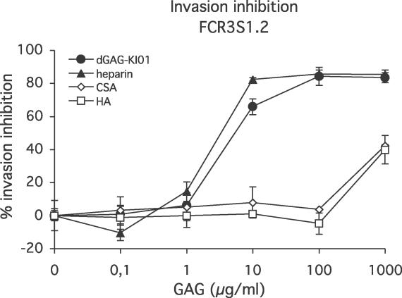
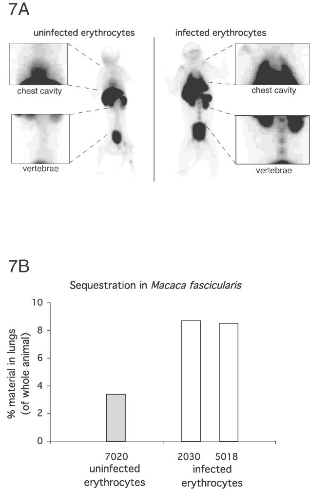
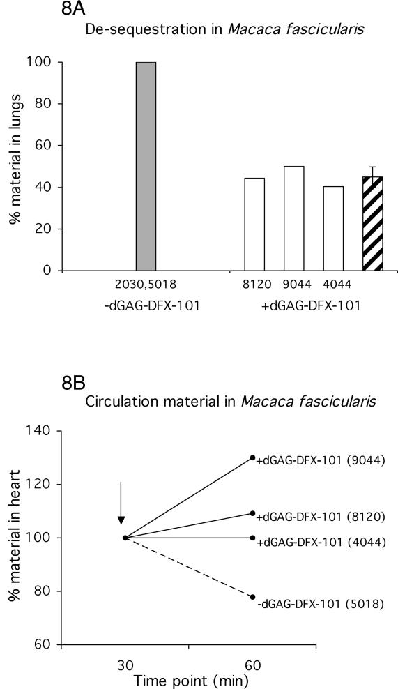
Severe human malaria is attributable to an excessive sequestration of Plasmodium falciparum-infected and uninfected erythrocytes in vital organs. Strains of P. falciparum that form rosettes and employ heparan sulfate as a host receptor are associated with development of severe forms of malaria. Heparin, which is similar to heparan sulfate in that it is composed of the same building blocks, was previously used in the treatment of severe malaria, but it was discontinued due to the occurrence of serious side effects such as intracranial bleedings. Here we report to have depolymerized heparin by periodate treatment to generate novel glycans (dGAG) that lack anticoagulant-activity. The dGAGs disrupt rosettes, inhibit merozoite invasion of erythrocytes and endothelial binding of P. falciparum-infected erythrocytes in vitro, and reduce sequestration in in vivo models of severe malaria. An intravenous injection of dGAGs blocks up to 80% of infected erythrocytes from binding in the micro-vasculature of the rat and releases already sequestered parasites into circulation. P. falciparum-infected human erythrocytes that sequester in the non-human primate Macaca fascicularis were similarly found to be released in to the circulation upon a single injection of 500 mug of dGAG. We suggest dGAGs to be promising candidates for adjunct therapy in severe malaria.


Conflict of interest statement

Competing interests. The authors declare competing financial interest. AMV, MW, and HPE hold stock and/or stock options in Dilafor AB, which owns the intellectual property of the substances generated in this manuscript. AMV has received research funding from Dilafor AB. The remaining authors have no known financial interest.

Figures

Figure 1
Figure 1. Binding of dGAG-KI01 to P. falciparum Antigens
(A) dGAG-KI01 binding to DBL1α was analyzed using an in-solution assay. A fixed concentration (4 μg/ml) was allowed to incubate with increasing concentrations of 3H-dGAG-KI01. Protein with bound dGAG was recovered by membrane filtration. (B) Aliquots of rosetting parasite cultures (UAS22, UAS29, UAS31, and FCR3S1.2) were incubated with 3H-dGAG-KI01. Cells were lysed in hypotonic buffer, and membranes (M) were separated from the supernatants (S). 3H-dGAG-KI01 bound to DBL1α and cell compartments were analyzed by scintillation counting.
Figure 2
Figure 2. Effect of Heparin and HS on P. falciparum Rosettes and Cytoadherence of IE In Vitro
(A) Aliquots of rosetting parasite cultures (FCR3S1.2) were treated with heparin or HS at different concentrations. The rosetting-rates were counted after 30 min incubation and compared with mock treated samples. The cytoadherence assays (B and C) with IE (FCR3S1.2) and heparin or HS were performed under orbital shaking (50 rpm) at 37 °C. Increasing concentrations of heparin and HS were added together with IE (B) or after letting the IE adhere (C). Unbound material was removed before the slides were fixed in glutaraldehyde, stained with Giemsa and analyzed by light microscopy at a x1,000 magnification.
Figure 3
Figure 3. Effect of dGAG-KI01 on P. falciparum Rosettes and Cytoadherence of IE In Vitro
(A) Aliquots of rosetting parasite cultures (UAS22, UAS29, UAS31, and FCR3S1.2) were treated with dGAG-KI01 at different concentrations. The rosetting rates were counted after 30-min incubation and compared with mock-treated samples. (B) and (C) For the cytoadherence assays, different P. falciparum IE (UAS22, UAS29, UAS31, and FCR3S1.2) were allowed to attach to rat lung sections under orbital shaking (50 rpm) at 37 °C. Different concentrations of dGAG-KI01 were added together with IE (B) or after letting the IE adhere (C). Unbound material was removed by washes before the slides were fixed in glutaraldehyde, stained with Giemsa, and analyzed by light microscopy at a 1,000× magnification.
Figure 4
Figure 4. Effects of Different GAGs on Reinvasion of P. falciparum Merozoites (FCR3S1.2) into Fresh Erythrocytes In Vitro
Parasite culture at throphozoite stage (≈25 h of development) at a 0.4% parasitemia and a 2% hematocrit were incubated with increasing concentrations of dGAG-KI01, heparin, CSA, or HA for 24–30 h at 37 °C. Parasitemias were estimated using FACS counting with a minimum of 50,000 cells per sample.
Figure 5
Figure 5. Injection of dGAG-KI01 into Rats Prevents Sequestration of P. falciparum (FCR3S1.2) IE in the Lungs
(A) and (B) Rats received either 99mTc-labeled IE simultaneously with different concentrations of the dGAG-KI01 (sequestration blocking) (A), or were first injected with 99mTc-labeled IE and, after three min, injected with dGAG-KI01 (de-sequestration) (B). (C) Whole-body summary images of rats captured 21–30 min after the injection of 99mTc-labeled IE alone (−dGAG-KI01) or when co-injected with 5 μg of dGAG-KI01 (+dGAG-KI01). (D) and (E) Rats were injected with 99mTc-labeled IE and subsequently with heparin (D) or the sulfated polysaccharide dextran sulfate (E). Rats were in all cases left in the gamma camera for 30 min after which the lungs were excised, measured for radioactivity, and compared to the level of radioactivity in the whole animal. Results are presented as relative amount in lungs compared with control animals receiving no dGAG-KI01, heparin, or dextran sulfate (control, adjusted to 100%; grey bars). White bars show the results of individual rats, and striped bars, the mean thereof.
Figure 6
Figure 6. Injection of dGAG-KI01 Decreases P. falciparum Sequestration of IE of Clinical Isolates in the Rat
(A) Rats were co-injected with 99mTc-labeled IE (clinical isolates: UAS22, UAS29, and UAS31) and 5 μg of the dGAG-KI01, or (B) rats were first administrated with 99mTc-labeled IE (clinical isolates: UAS22, UAS29, and UAS31) and after 3 min, injected with different concentrations of the dGAG-KI01. Rats were in all cases left in the gamma camera for 30 min, after which the lungs were excised, measured for radioactivity, and compared to the level of radioactivity in the whole animal. Results are presented as relative amount in lungs compared with control animal receiving no dGAG-KI01 (control, adjusted to 100%; grey bars). White bars show the results of individual rats, and the striped bars, the mean thereof.
Figure 7
Figure 7. Sequestration of P. falciparum IE of Parasite FCR3S1.2 in Macaca fascicularis
Monkeys were injected with 99mTc-labeled IE of FCR3S1.2 or with 99mTc-labeled uninfected human erythrocytes and left for 60 min followed by analysis in an X-ray–equipped triple-headed gamma camera (ID 7020, uninfected erythrocytes; ID 2030 and 5018, IE). (A) Shows the whole-body summary images captured 60–75 min after the injection of the 99mTc-labeled cells. Amplified areas show the chest cavities and the vertebrae. The accumulation of 99mTc-labeled IE in the lungs and in the bone marrow suggests their specific sequestration. The activity in the heart of the animal that received uninfected erythrocytes indicates that the erythrocytes are in circulation. (B) Shows the relative amount of material in the lungs as compared with the activity of the whole animal and measured for 15 min (60 to 75 min after injection). White bars show results of individual monkeys injected with IE, whereas the grey bars show the results of the control animal injected with uninfected human erythrocytes.
Figure 8
Figure 8. Injection of dGAG-DFX-101 Releases Sequestered P. falciparum IE into Circulation in M. fascicularis
Five macaques (M. fascicularis) were injected with 99mTc-labeled IE of the parasite FCR3S1.2 and left for 30 min. Three of the monkeys (ID 8120, 9044, and 4044) each received 500-μg dGAG-DFX-101 by intravenous injection while two monkeys (ID 2030 and 5018) were left untreated. (A) Shows the level of material in the lungs. The dGAG was injected at time point 30 min and the radioactivity in the animals were measured for 15 min 60 to 75 min after injection. The level of radioactive material in the lungs was compared with the amount of radioactive material in the whole animal. Results are presented as relative amount in lungs compared with control animal receiving no dGAG-DFX-101 (control, adjusted to 100%; grey bar). White bars show the relative amount of material in individual monkeys, and the striped bars, the mean thereof. (B) Shows the level of material in the hearts before and after the injection of the dGAG-DFX-101. The radioactive material in the hearts (i.e., in circulation) increases (9%, ID 8120; 30% ID 9044; and 0% ID 4044) after treatment with dGAG-DFX-101. In contrast, the amount of material in the heart of the monkey left untreated decreased by 22% (ID 5018). The analysis of the radioactivity in the hearts was performed by comparing amounts of material present at time point 30 min and at time point 60 min, after the dGAG-DFX-101 was injected. Time point 30 min was adjusted to 100%. The arrow shows the time point of injection of the dGAG-DFX-101.

References

    1. MacPherson GG, Warrell MJ, White NJ, Looareesuwan S, Warrell DA. Human cerebral malaria. A quantitative ultrastructural analysis of parasitized erythrocyte sequestration. Am J Pathol. 1985;119:385–401. - PMC - PubMed
    1. Carlson J, Helmby H, Hill AV, Brewster D, Greenwood BM, et al. Human cerebral malaria: Association with erythrocyte rosetting and lack of anti-rosetting antibodies. Lancet. 1990;336:1457–1460. - PubMed
    1. Rowe A, Obeiro J, Newbold CI, Marsh K. Plasmodium falciparum rosetting is associated with malaria severity in Kenya. Infect Immun. 1995;63:2323–2326. - PMC - PubMed
    1. Heddini A, Pettersson F, Kai O, Shafi J, Obiero J, et al. Fresh isolates from children with severe Plasmodium falciparum malaria bind to multiple receptors. Infect Immun. 2001;69:5849–5856. - PMC - PubMed
    1. Roberts DJ, Pain A, Kai O, Kortok M, Marsh K. Autoagglutination of malaria-infected red blood cells and malaria severity. Lancet. 2000;355:1427–1428. - PubMed

Publication types

MeSH terms

Substances