. 2006 Nov 24;364(2):152-69.
doi: 10.1016/j.jmb.2006.08.080. Epub 2006 Sep 1.

Substrate recognition, protein dynamics, and iron-sulfur cluster in Pseudomonas aeruginosa adenosine 5'-phosphosulfate reductase

Affiliations

Substrate recognition, protein dynamics, and iron-sulfur cluster in Pseudomonas aeruginosa adenosine 5'-phosphosulfate reductase

Justin Chartron et al. J Mol Biol. .

Abstract

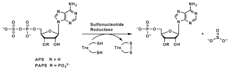
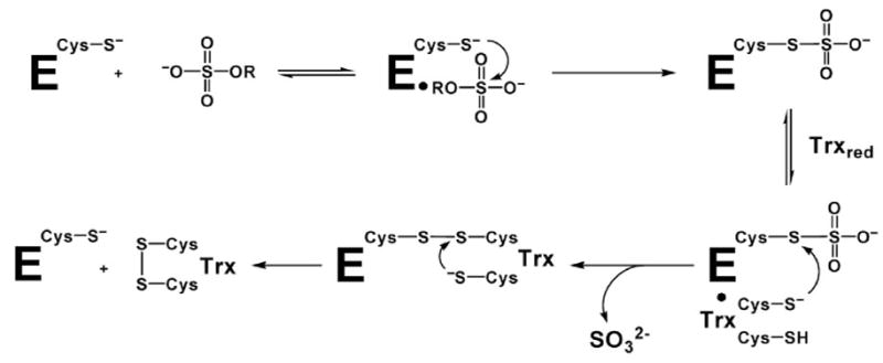
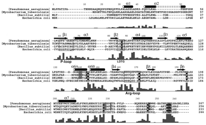
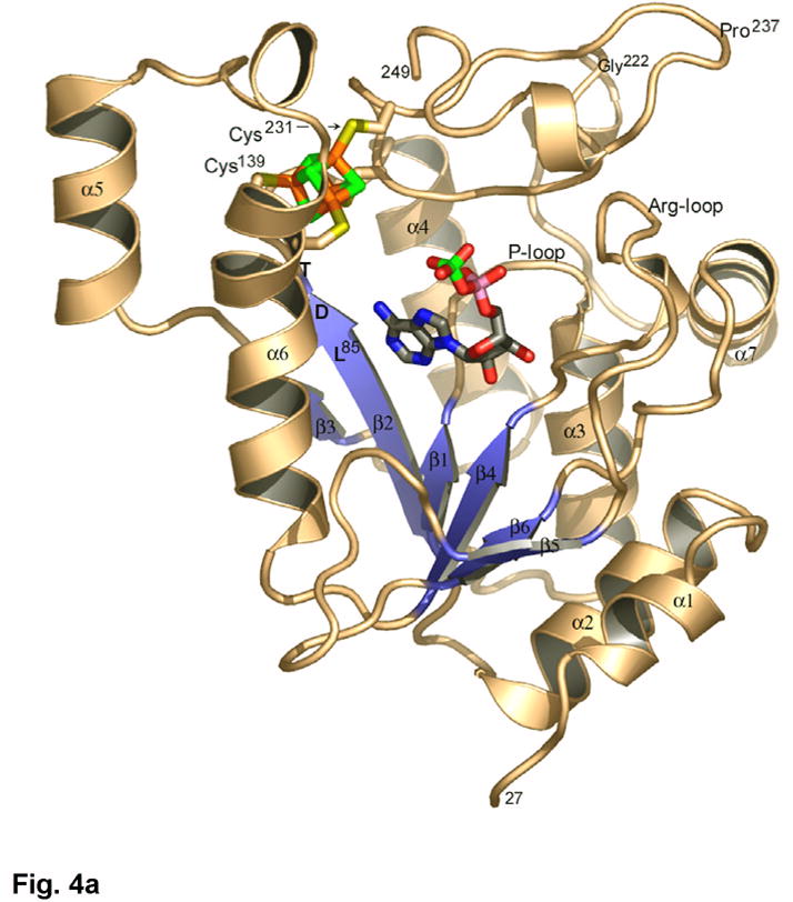
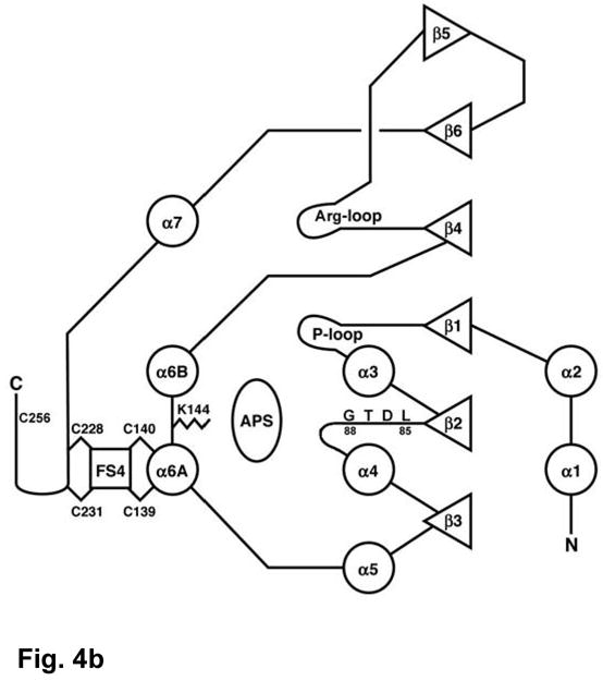
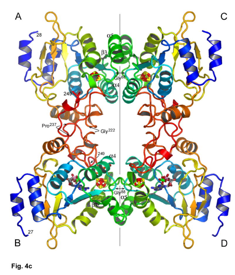
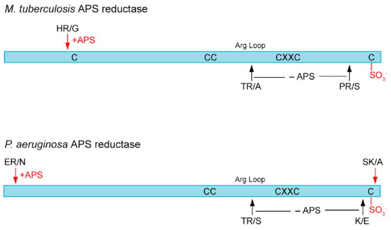
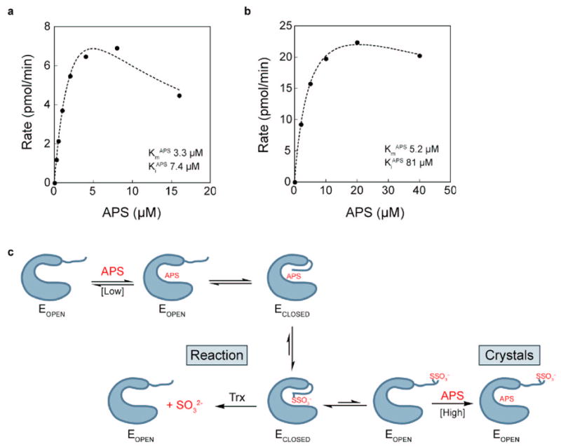
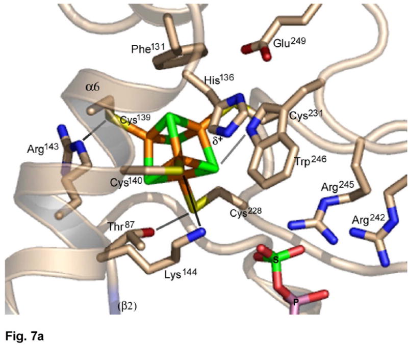
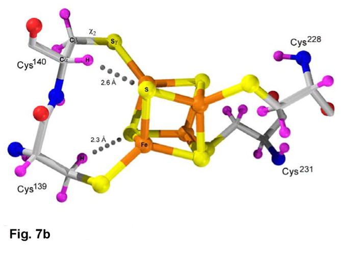
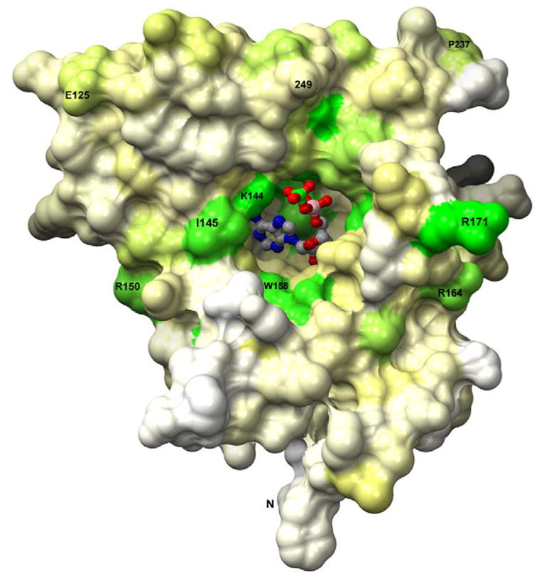
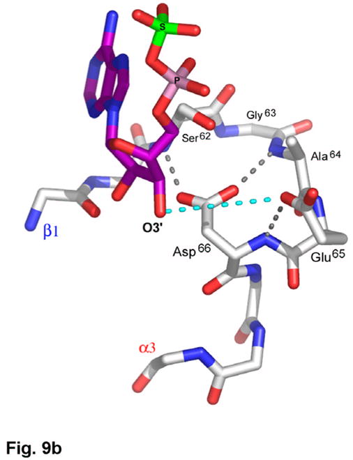
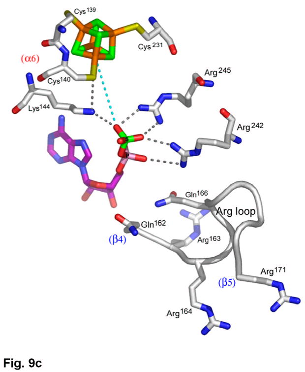
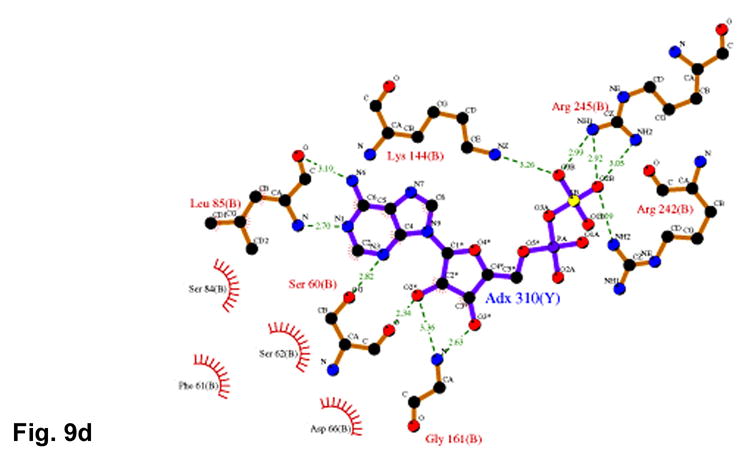
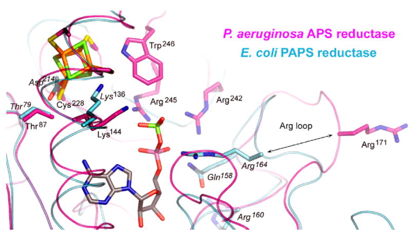
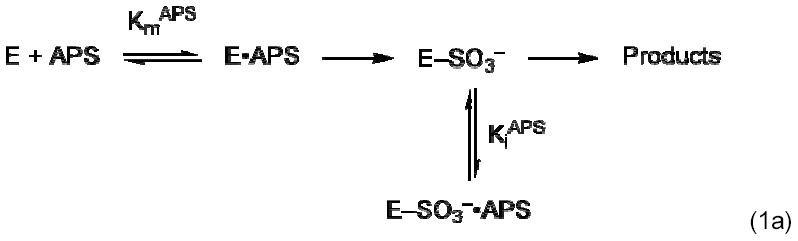
APS reductase catalyzes the first committed step of reductive sulfate assimilation in pathogenic bacteria, including Mycobacterium tuberculosis, and is a promising target for drug development. We report the 2.7 A resolution crystal structure of Pseudomonas aeruginosa APS reductase in the thiosulfonate intermediate form of the catalytic cycle and with substrate bound. The structure, high-resolution Fourier transform ion cyclotron resonance (FT-ICR) mass spectrometry, and quantitative kinetic analysis, establish that the two chemically discrete steps of the overall reaction take place at distinct sites on the enzyme, mediated via conformational flexibility of the C-terminal 18 residues. The results address the mechanism by which sulfonucleotide reductases protect the covalent but labile enzyme-intermediate before release of sulfite by the protein cofactor thioredoxin. P. aeruginosa APS reductase contains an [4Fe-4S] cluster that is essential for catalysis. The structure reveals an unusual mode of cluster coordination by tandem cysteine residues and suggests how this arrangement might facilitate conformational change and cluster interaction with the substrate. Assimilatory 3'-phosphoadenosine 5'-phosphosulfate (PAPS) reductases are evolutionarily related, homologous enzymes that catalyze the same overall reaction, but do so in the absence of an [Fe-S] cluster. The APS reductase structure reveals adaptive use of a phosphate-binding loop for recognition of the APS O3' hydroxyl group, or the PAPS 3'-phosphate group.


Figures

Figure 1
Figure 1
Reaction catalyzed by sulfonucleotide reductases.
Figure 2
Figure 2
Mechanism of sulfonucleotide reduction.
Figure 3
Figure 3
Primary sequences for APS reductases from P. aeruginosa, M. tuberculosis, and B. subtilis, and for PAPS reductase from E. coli, are shown based on alignment of 38 APS and 34 PAPS reductases (Supplementary Figure 2). Secondary structure elements are denoted for P. aeruginosa APS reductase, and residues within the P-loop (60–66), LDTG motif (85–88), and Arg-loop (163–171), are boxed. The bar graph indicates the degree of conservation based on all 72 sequences (Supplementary Figure 2). Strictly conserved residues are outlined in black; six additional residues conserved in 38 APS reductases that ligate or interact with the [4Fe-4S] cluster are boxed in gray.
Figure 4
Figure 4
(a) The structure of P. aeruginosa APS reductase comprises a central 6-stranded β sheet flanked by α-helices and loops that create a deep active site cavity. The substrate APS binds across the C-terminal ends of the β-strands, and occupies the active site in half of the independent copies of the protein in the crystal (subunit B is shown). The [4Fe-4S] cluster is ligated by four cysteines at residues 139 and 140 on the long, kinked helix α6, and at residues 228 and 231 at the tip of a β-turn. (b) Secondary structure topology of P. aeruginosa APS reductase indicating α-helix and β-strand elements, active site loops, conserved residues Lys144 and Cys256, the LDTG motif, and Cys ligand connectivity to the [4Fe-4S] cluster. (c) Triclinic crystals of P. aeruginosa APS reductase contain two tetramers in the asymmetric unit. Each chain of the ABCD tetramer is colored blue->red from N- to C-terminus (residues 27–249). Labeled residues are involved in inter-subunit contacts.
Figure 4
Figure 4
(a) The structure of P. aeruginosa APS reductase comprises a central 6-stranded β sheet flanked by α-helices and loops that create a deep active site cavity. The substrate APS binds across the C-terminal ends of the β-strands, and occupies the active site in half of the independent copies of the protein in the crystal (subunit B is shown). The [4Fe-4S] cluster is ligated by four cysteines at residues 139 and 140 on the long, kinked helix α6, and at residues 228 and 231 at the tip of a β-turn. (b) Secondary structure topology of P. aeruginosa APS reductase indicating α-helix and β-strand elements, active site loops, conserved residues Lys144 and Cys256, the LDTG motif, and Cys ligand connectivity to the [4Fe-4S] cluster. (c) Triclinic crystals of P. aeruginosa APS reductase contain two tetramers in the asymmetric unit. Each chain of the ABCD tetramer is colored blue->red from N- to C-terminus (residues 27–249). Labeled residues are involved in inter-subunit contacts.
Figure 5
Figure 5
Sites of trypsin proteolysis in M. tuberculosis and P. aeruginosa APS reductase. In the absence of substrate (-APS, black arrows) both the Arg-loop and the C-terminal tail are cleaved; in the presence of equimolar substrate (+APS, red arrows), formation of the thiosulfonate intermediate protects these structural elements, and only sites at the extreme N- and C-termini are cleaved. See Supplementary Figure 5 for SDS-PAGE analysis and peptide masses.
Figure 6
Figure 6
Dependence of APS reductase activity on APS concentration. (a) Reaction rate measured in the presence of 1 μM thioredoxin. (b) Reaction rate measured in the presence of 10 μM thioredoxin. Data were fit with Eq. 1b, derived from the inhibitory model in Eq. 1a (see Methods). (c) Model for mobility of C-terminal tail during APS reduction. The C-terminal peptide of APS reductase (E) can be in an open or closed conformation. Higher concentrations of Trx favor sulfite release and regeneration of free enzyme, resulting in a higher apparent KiAPS. In the presence of excess APS, but absence of Trx, the enzyme is trapped in the inhibitory open form, as observed in the crystals.
Figure 7
Figure 7
(a) The environment of the [4Fe-4S] cluster in P. aeruginosa APS reductase. The [4Fe-4S] cluster is ligated by four cysteines at residues 139, 140, 228 and 231. Four conserved residues participate in charged or polar NH…S or OH…S hydrogen bonds to inorganic S or cysteine Sγ atoms – Thr87, Arg143, Lys144 and Trp246. In addition, His136 may be hydrogen bonded to Cys140. (b) Molecular details of the [4Fe-4S] cluster and its cysteine ligands in P. aeruginosa APS reductase. The positions of H atoms, calculated based on C and N atomic coordinates, indicate steric clashes (dotted lines). Due to linkage of tandem cysteines in an α-helix, the χ2 torsion angle is cis (Supplementary Figure 6) and the Cys140 Cα-H atom is ~2.6 Å from inorganic S of the cluster. In addition, an H atom on Cβ of Cys139 is ~2.3 Å from a S atom. The sum of van der Waals radii for S and H is 3.00 Å.
Figure 7
Figure 7
(a) The environment of the [4Fe-4S] cluster in P. aeruginosa APS reductase. The [4Fe-4S] cluster is ligated by four cysteines at residues 139, 140, 228 and 231. Four conserved residues participate in charged or polar NH…S or OH…S hydrogen bonds to inorganic S or cysteine Sγ atoms – Thr87, Arg143, Lys144 and Trp246. In addition, His136 may be hydrogen bonded to Cys140. (b) Molecular details of the [4Fe-4S] cluster and its cysteine ligands in P. aeruginosa APS reductase. The positions of H atoms, calculated based on C and N atomic coordinates, indicate steric clashes (dotted lines). Due to linkage of tandem cysteines in an α-helix, the χ2 torsion angle is cis (Supplementary Figure 6) and the Cys140 Cα-H atom is ~2.6 Å from inorganic S of the cluster. In addition, an H atom on Cβ of Cys139 is ~2.3 Å from a S atom. The sum of van der Waals radii for S and H is 3.00 Å.
Figure 8
Figure 8
APS binds in a deep active site pocket of P. aeruginosa APS reductase such that only the sulfate group is exposed. The solvent accessible surface is depicted, and residues are colored by degree of sequence conservation (Supplementary Figure 2) among 72 APS and PAPS reductases; green – most conserved; yellow – partial conservation; white – variable). The orientation is similar to that in Figure 4(a).
Figure 9
Figure 9
(a) Recognition of the adenosine moiety of APS by conserved residues on strands β1, β2, and β4, which participate in four main chain hydrogen bonds with adenine and the ribose O2′ hydroxyl group. In addition, these residues stabilize the conformation of the β-strands through hydrogen bonds within two conserved motifs (Leu85Asp86Thr87Gly88 and Thr160Gly161). (b) The P-loop comprising conserved residues 60–66 connects strand β1 and helix α3 in the P. aeruginosa APS reductase active site. Three amides hydrogen bond with Glu65 or Asp66, but in PAPS reductases these acidic residues are replaced by Gln and Ala or Ser, respectively. The distance between Glu65 and the O3′ of ribose is 5.3 Å (cyan dotted line). (c) Conserved basic residues in the vicinity of the active site interact with the phosphate and sulfate groups of APS, or reside on the Arg-loop, comprising residues 162–175 between strands β4 and β5. The shortest distance between a sulfate oxygen atom and Fe of the [4Fe-4S] cluster is ~7.0 Å (cyan dotted line). (d) Summary of all active site contacts to APS in subunit B of the asymmetric unit (PDB deposition 2GOY) plotted in two dimensions; hydrogen bond distances are indicated in Å.
Figure 9
Figure 9
(a) Recognition of the adenosine moiety of APS by conserved residues on strands β1, β2, and β4, which participate in four main chain hydrogen bonds with adenine and the ribose O2′ hydroxyl group. In addition, these residues stabilize the conformation of the β-strands through hydrogen bonds within two conserved motifs (Leu85Asp86Thr87Gly88 and Thr160Gly161). (b) The P-loop comprising conserved residues 60–66 connects strand β1 and helix α3 in the P. aeruginosa APS reductase active site. Three amides hydrogen bond with Glu65 or Asp66, but in PAPS reductases these acidic residues are replaced by Gln and Ala or Ser, respectively. The distance between Glu65 and the O3′ of ribose is 5.3 Å (cyan dotted line). (c) Conserved basic residues in the vicinity of the active site interact with the phosphate and sulfate groups of APS, or reside on the Arg-loop, comprising residues 162–175 between strands β4 and β5. The shortest distance between a sulfate oxygen atom and Fe of the [4Fe-4S] cluster is ~7.0 Å (cyan dotted line). (d) Summary of all active site contacts to APS in subunit B of the asymmetric unit (PDB deposition 2GOY) plotted in two dimensions; hydrogen bond distances are indicated in Å.
Figure 10
Figure 10
Superposition of the structures of P. aeruginosa APS reductase and E. coli PAPS reductase showing the positions of the [4Fe-4S] cluster and APS bound in APS reductase with respect to conserved active site residues in both enzymes. Distinct conformations of the Arg loop are indicated, showing that the loop folds into the active site of PAPS reductase in the absence of substrate. Arg164 (P. aeruginosa) and Arg171 (E. coli) are equivalent residues, conserved in 72 species of APS and PAPS reductases (Supplementary Figure 2).
None

References

    1. Bick JA, Dennis JJ, Zylstra GJ, Nowack J, Leustek T. Identification of a new class of 5′-adenylylsulfate (APS) reductases from sulfate-assimilating bacteria. J Bacteriol. 2000;182:135–142. - PMC - PubMed
    1. Kopriva S, Buchert T, Fritz G, Suter M, Weber M, Benda R, Schaller J, Feller U, Schurmann P, Schunemann V, Trautwein AX, Kroneck PM, Brunold C. Plant adenosine 5′-phosphosulfate reductase is a novel iron-sulfur protein. J Biol Chem. 2001;276:42881–42886. - PubMed
    1. Williams SJ, Senaratne RH, Mougous JD, Riley LW, Bertozzi CR. 5′-Adenosinephosphosulfate lies at a metabolic branch point in mycobacteria. J Biol Chem. 2002;277:32606–32615. - PubMed
    1. Leyh TS, Taylor JC, Markham GD. The sulfate activation locus of Escherichia coli K12: cloning, genetic, and enzymatic characterization. J Biol Chem. 1988;263:2409–2416. - PubMed
    1. Mougous JD, Lee DH, Hubbard SC, Schelle MW, Vocadlo DJ, Berger JM, Bertozzi CR. Molecular basis for G protein control of the prokaryotic ATP sulfurylase. Mol Cell. 2006;21:109–122. - PubMed

Publication types

Associated data