Protection against polyoma virus-induced tumors is perforin-independent
- PMID: 17011010
- PMCID: PMC2861337
- DOI: 10.1016/j.virol.2006.08.044
Protection against polyoma virus-induced tumors is perforin-independent
Abstract
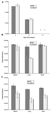
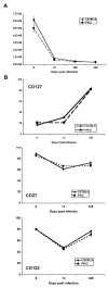
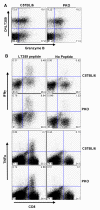
CD8 T cells are necessary for controlling tumors induced by mouse polyoma virus (PyV), but the effector mechanism(s) responsible have not been determined. We examined the PyV tumorigenicity in C57BL/6 mice mutated in Fas or carrying targeted disruptions in the perforin gene or in both TNF receptor type I and type II genes. Surprisingly, none of these mice developed tumors. Perforin/Fas double-deficient radiation bone marrow chimeric mice were also resistant to PyV-induced tumors. Anti-PyV CD8 T cells in perforin-deficient mice were found not to differ from wild type mice with respect to phenotype, capacity to produce cytokines or maintenance of memory T cells, indicating that perforin does not modulate the PyV-specific CD8 T cell response. In addition, virus was cleared and persisted to similar extents in wild type and perforin-deficient mice. In summary, perforin/granzyme exocytosis is not an essential effector pathway for protection against PyV infection or tumorigenesis.
Figures


Similar articles
-
Immunity to polyomavirus infection: the polyomavirus-mouse model.Semin Cancer Biol. 2009 Aug;19(4):244-51. doi: 10.1016/j.semcancer.2009.02.003. Epub 2009 Feb 14. Semin Cancer Biol. 2009. PMID: 19505652 Free PMC article. Review.
-
Costimulation requirements for antiviral CD8+ T cells differ for acute and persistent phases of polyoma virus infection.J Immunol. 2006 Feb 1;176(3):1814-24. doi: 10.4049/jimmunol.176.3.1814. J Immunol. 2006. PMID: 16424212
-
T cell-independent antibody-mediated clearance of polyoma virus in T cell-deficient mice.J Exp Med. 1996 Feb 1;183(2):403-11. doi: 10.1084/jem.183.2.403. J Exp Med. 1996. PMID: 8627153 Free PMC article.
-
Generation of antiviral major histocompatibility complex class I-restricted T cells in the absence of CD8 coreceptors.J Virol. 2008 May;82(10):4697-705. doi: 10.1128/JVI.02698-07. Epub 2008 Mar 12. J Virol. 2008. PMID: 18337581 Free PMC article.
-
Immunity to polyoma virus infection and tumorigenesis.Viral Immunol. 2001;14(3):199-216. doi: 10.1089/088282401753266738. Viral Immunol. 2001. PMID: 11572632 Review.
Cited by
-
Immunity to polyomavirus infection: the polyomavirus-mouse model.Semin Cancer Biol. 2009 Aug;19(4):244-51. doi: 10.1016/j.semcancer.2009.02.003. Epub 2009 Feb 14. Semin Cancer Biol. 2009. PMID: 19505652 Free PMC article. Review.
-
T cell deficiency precipitates antibody evasion and emergence of neurovirulent polyomavirus.Elife. 2022 Nov 7;11:e83030. doi: 10.7554/eLife.83030. Elife. 2022. PMID: 36341713 Free PMC article.
-
Gamma interferon controls mouse polyomavirus infection in vivo.J Virol. 2011 Oct;85(19):10126-34. doi: 10.1128/JVI.00761-11. Epub 2011 Jul 20. J Virol. 2011. PMID: 21775464 Free PMC article.
-
Persistence of viral infection despite similar killing efficacy of antiviral CD8(+) T cells during acute and chronic phases of infection.Virology. 2010 Sep 15;405(1):193-200. doi: 10.1016/j.virol.2010.05.029. Epub 2010 Jun 26. Virology. 2010. PMID: 20580390 Free PMC article.
-
Allogeneic differences in the dependence on CD4+ T-cell help for virus-specific CD8+ T-cell differentiation.J Virol. 2007 Dec;81(24):13743-53. doi: 10.1128/JVI.01778-07. Epub 2007 Oct 3. J Virol. 2007. PMID: 17913814 Free PMC article.
References
-
- Allison AC, Monga JN, Hammond V. Increased susceptibility to virus oncogenesis of congenitally thymus-deprived nude mice. Nature. 1974;252(5485):746–747. - PubMed
-
- Badovinac VP, Hamilton SE, Harty JT. Viral infection results in massive CD8+ T cell expansion and mortality in vaccinated perforin-deficient mice. Immunity. 2003;18(4):463–474. - PubMed
-
- Badovinac VP, Tvinnereim AR, Harty JT. Regulation of antigen-specific CD8+ T cell homeostasis by perforin and interferon-γ. Science. 2000;290(5495):1354–1358. - PubMed
-
- Barber DL, Wherry EJ, Ahmed R. Cutting edge: rapid in vivo killing by memory CD8 T cells. J. Immunol. 2003;171(1):27–31. - PubMed
-
- Berebbi M, Dandolo L, Hassoun J, Bernard AM, Blangy D. Specific tissue targeting of polyoma virus oncogenicity in athymic nude mice. Oncogene. 1988;2(2):149–156. - PubMed
Publication types
MeSH terms
Substances
Grants and funding
LinkOut - more resources
Full Text Sources
Research Materials
Miscellaneous

