. 2006;34(19):e128.
doi: 10.1093/nar/gkl679. Epub 2006 Sep 29.

A novel endonuclease IV post-PCR genotyping system

Affiliations

A novel endonuclease IV post-PCR genotyping system

Igor V Kutyavin et al. Nucleic Acids Res. 2006.

Abstract

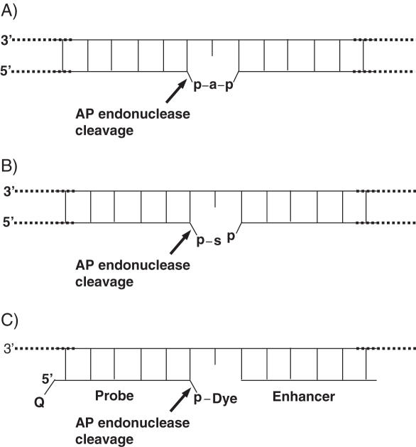
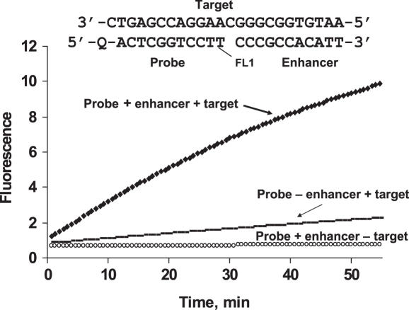
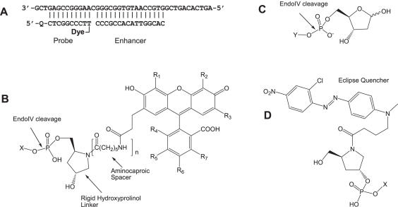
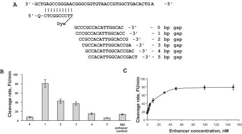
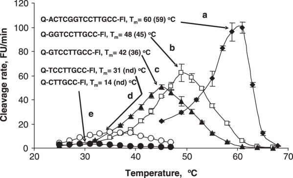
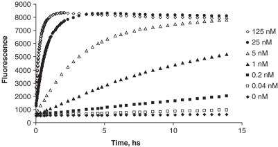
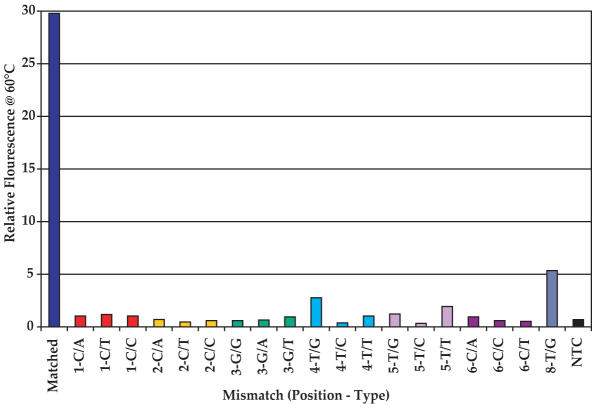
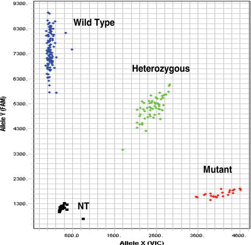
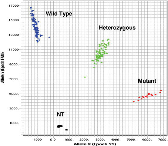
Here we describe a novel endonuclease IV (Endo IV) based assay utilizing a substrate that mimics the abasic lesions that normally occur in double-stranded DNA. The three component substrate is characterized by single-stranded DNA target, an oligonucleotide probe, separated from a helper oligonucleotide by a one base gap. The oligonucleotide probe contains a non-fluorescent quencher at the 5' end and fluorophore attached to the 3' end through a special rigid linker. Fluorescence of the oligonucleotide probe is efficiently quenched by the interaction of terminal dye and quencher when not hybridized. Upon hybridization of the oligonucleotide probe and helper probe to their complementary target, the phosphodiester linkage between the rigid linker and the 3' end of the probe is efficiently cleaved, generating a fluorescent signal. In this study, the use of the Endo IV assay as a post-PCR amplification detection system is demonstrated. High sensitivity and specificity are illustrated using single nucleotide polymorphism detection.


Figures

Figure 1
Figure 1
(A) First double strand DNA lesion where ‘a’ represents an abasic 2′-deoxyribose residue and ‘p’ is a phosphate group. (B) Second DNA lesion, formed by spontaneous or enzymatic cleavage of the phosphodiester bond between the 3′-hydroxyl group of the 2′-deoxyribose (abasic site) and the nearest nucleotide of the DNA strand. ‘s’ represents several products of 2′-deoxyribose degradation. (C) Artificial abasic lesion generated by the novel probe and enhancer. Arrows indicated the specific cleavage by AP endonuclease.
Scheme 1
Scheme 1
Synthesis of methyl 3-(3-chloro-2,4-dihydroxyphenyl)propanoate (4a) required for the preparation of PFP esters 11(a and c).
Scheme 2
Scheme 2
Synthesis of the pentafluorophenyl esters 11(ac) required for the preparation of dye-modified CPG supports.
Scheme 3
Scheme 3
Synthesis of the dye-modified CPG supports 12(ac).
Figure 2
Figure 2
Monitoring of fluorescence by the Endo IV cleavage reaction was performed in 5 mM MgCl2, 20 mM Tris–HCl (pH 8.5). The three component substrate complex used in these experiments is shown. FL1 is a fluorescein derivative and Q is the Eclipse® Dark Quencher, structures of which are shown in Figure 3B and D, respectively. The reaction mixture contained Endo IV at 0.04 U/μl concentration, probe and enhancer at 150 nM, and the target at 5 nM. The experiment was performed on LightCycler.
Figure 3
Figure 3
(A) Endonuclease IV model assay showing a target sequence, a 16mer enhancer (calculated Tm 50°C) and a 10mer Endo IV probe (calculated Tm 46.7°C). Q is Eclipse Dark Quencher and the Dyes are compounds from FL1 to FL4. (B) Dyes with hydroxyprolinol (n = 0) and extended (n = 1) hydroxyprolinol linkers. (C) Natural abasic substrate. (D) Eclipse Dark Quencher attached to the 5′ end of the probe. X shows the connection to the oligonucleotide probe. Y shows the connection to the single-stranded DNA.
Figure 4
Figure 4
(A) Endo IV assay investigating a gap of 0–5 bases between the probe and primer. (B) Shows the cleavage rate dependence on the gap between the probe and primer in reference to the cleavage rate when no enhancer is present. (C) Shows the cleavage rate in the presence of different concentrations of a 16mer enhancer. Experiments were performed in quintiplicate.
Figure 5
Figure 5
The effect of temperature on the cleavage rate of probes with different length and calculated Tm. The numbers in parentheses are the determined Tms. ‘nd’ is not determined. The target and enhancers sequences are, respectively, 5′-AGTCACAGTCGGTGCCAATGTGGCGGGCAAGGACCGAGTCG-3′ and 3′-CAGCCACGGTTACACCG-5′.
Figure 6
Figure 6
Dependence of rates of fluorescence generation on target concentration. Probe, enhancer and target sequences are shown in Figure 3. The concentration of reagents were 150 nM probe, 150 nM enhancer, 0.2 U/μl enzyme, 5 mM MgCl2 in 20 mM Tris–HCl buffer, pH 8.5. The reaction was performed at 45°C.
Figure 7
Figure 7
The comparison of the change in relative signal fluorescence of match and different mismatches at different positions in a 14mer probe in an Endo IV assay run at 60°C. The probe sequence of the matched probe and target sequence are, respectively, 5′-Q-ACTCGGTCCTTGCC-FL-3′ and 5′-AGTCACAGTCGGTGCCAATGTGGCGGGCAAGGACCGAGTCG-3′. All the complementary sequences indicating the different mismatches are shown in the Supplementary Data. NTC is the no template control.
Figure 8
Figure 8
A scatter plot of the genotyping of an agouti signal protein (ASIP-01) mutation. The probe sequences for the wild-type and mutant alleles are, respectively, 5′-Q-ACCTTCATTCCC-FL1-3′ and 5′-Q-GACCTTCATTCTC-FL3-3′. The mismatch is underlined. NT is no template control and Q is the Eclipse Quencher. The primer sequences are TCCAGTGCCCCTCATATTTTCC and CTCCTGTCAACAGTAACAAACTTC.
Figure 9
Figure 9
A scatter plot of the genotyping of an adenomatous polyposis coli (APC-03) mutation. The probe sequences for the wild-type and mutant alleles are, respectively, 5′-Q-TACTTTCTGTGGC-FL1-3′ and 5′-Q-CTTTTCCGTGGC-FL3-3′. The mismatch is underlined. NT is no template control and Q is the Eclipse Dark Quencher. Primer sequences are AGCACTCAGGCTGGATGAACAA and GTTCAGAGGGTCCAGGTTCTT.

References

    1. Holland P.M., Abramson R.D., Watson R., Gelfand D.H. Detection of specific polymerase chain reaction product by utilizing the 5′→3′ exonuclease activity of Thermus aquaticus. Proc. Natl Acad. Sci., USA. 1991;88:7276–7280. - PMC - PubMed
    1. Livak K.J. Allelic discrimination using fluorogenic probes and the 5′ nuclease assay. Gen. Anal. 1999;14:143–149. - PubMed
    1. Tang Y.-W., Procop G.W., Persing D.H. Molecular diagnostics of infectious diseases. Clin. Chem. 1997;43:2021–2038. - PubMed
    1. Livak K.J., Flood S.J., Marmaro J., Giusti W., Deetz K. Oligonucleotides with fluorescent dyes at opposite ends provide a quenched probe system useful for detecting PCR product and nucleic acid hybridization. PCR Methods Appl. 1995;4:357–362. - PubMed
    1. Marras S.A., Kramer F.R., Tyagi S. Efficiencies of fluorescence resonance energy transfer and contact-mediated quenching in oligonucleotide probes. Nucleic Acids Res. 2002;30:e122. - PMC - PubMed

Publication types

MeSH terms

Substances