Review
. 2006;34(19):5402-15.
doi: 10.1093/nar/gkl655. Epub 2006 Sep 29.

Quadruplex DNA: sequence, topology and structure

Affiliations
Review

Quadruplex DNA: sequence, topology and structure

Sarah Burge et al. Nucleic Acids Res. 2006.

Abstract

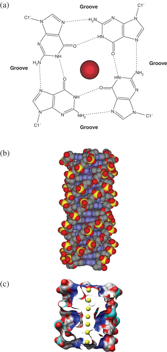
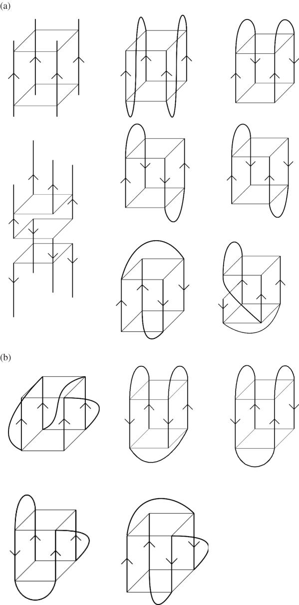
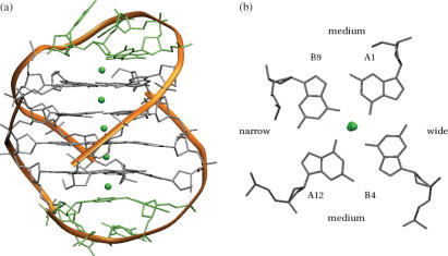
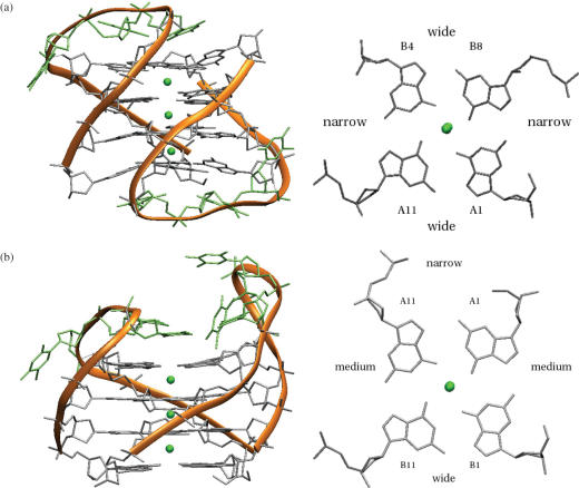
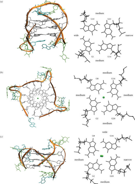
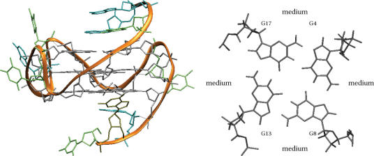
G-quadruplexes are higher-order DNA and RNA structures formed from G-rich sequences that are built around tetrads of hydrogen-bonded guanine bases. Potential quadruplex sequences have been identified in G-rich eukaryotic telomeres, and more recently in non-telomeric genomic DNA, e.g. in nuclease-hypersensitive promoter regions. The natural role and biological validation of these structures is starting to be explored, and there is particular interest in them as targets for therapeutic intervention. This survey focuses on the folding and structural features on quadruplexes formed from telomeric and non-telomeric DNA sequences, and examines fundamental aspects of topology and the emerging relationships with sequence. Emphasis is placed on information from the high-resolution methods of X-ray crystallography and NMR, and their scope and current limitations are discussed. Such information, together with biological insights, will be important for the discovery of drugs targeting quadruplexes from particular genes.


Figures

Figure 1
Figure 1
(a) The arrangement of guanine bases in the G-quartet, shown together with a centrally placed metal ion. Hydrogen bonds are shown as dotted lines, and the positions of the grooves are indicated. (b) The poly(dG) four-fold, right-handed helix. (c) Surface view representation of a quadruplex structure comprising eight G-quartets, with the central channel exposed to show an array of metal ions (coloured yellow).
Figure 2
Figure 2
(a) Some possible topologies for simple tetramolecular (on the left-hand side) and bimolecular quadruplexes. Strand polarities are shown by arrows. (b) Some possible topologies for simple unimolecular quadruplexes.
Figure 3
Figure 3
The crystal structure (28) of the bimolecular quadruplex formed by the O.nova telomeric sequence d(G4T4G4) (PDB entry 1JPQ). (a) Overall topology is indicated by the ribbon representation in orange. The details of the molecular structure are also shown. Potassium ions are shown as green spheres. (b) A projection down the central channel, indicating the relative widths of the four grooves
Figure 4
Figure 4
Crystal structure (26) of the two bimolecular quadruplexes found in the crystal structure of d(G4T3G4) (a) two views of the head-to-tail quadruplex (PDB entry 2AVH). (b) Two views of the head-to-head quadruplex (PDB entry 2AVJ).
Figure 5
Figure 5
Structures of the human unimolecular telomeric quadruplex formed from the sequence d[AGGG(TTAGGG)3]. In each case two views are shown (a) one of the deposited structures of the Na+ form, determined by NMR (PDB entry 143D) (54), with a diagonal and two lateral loops. (b) K+ form A, determined by crystallography (PDB entry 1KF1) (23), with three strand-reversal loops (c) K+ form B, showing the topology determined by NMR (75,96), with one strand-reversal and two lateral loops. Nucleotide loop conformations for the detailed atomic structure shown here have been obtained from a molecular dynamics simulation performed by Sarah Burge that has used this topology as a starting-point. The NMR-derived structure of one of the sequences determined experimentally (96), is also available as PDB entry id 2KGU.
Figure 6
Figure 6
NMR-derived topology and one of the deposited structures of the c-myc quadruplex (86) (PDB entry 1XAV).

References

    1. Gellert M., Lipsett M.N., Davies D.R. Helix formation by guanylic acid. Proc. Natl Acad. Sci. USA. 1962;48:2013–2018. - PMC - PubMed
    1. Zimmerman S.B., Cohen G.H., Davies D.R. X-ray fiber diffraction and model-building study of polyguanylic acid and polyinosinic acid. J. Mol. Biol. 1975;92:181–192. - PubMed
    1. Arnott S., Chandrasekaran R., Marttila C.M. Structures for polyinosinic acid and polyguanylic acid. Biochem. J. 1974;141:537–543. - PMC - PubMed
    1. Howard F.B., Frazier J., Miles H.T. Stable and metastable forms of poly(G) Biopolymers. 1977;16:791–809. - PubMed
    1. Williamson J.R. G-tetrad structures in telomeric DNA. Ann. Rev. Biophys. Biomol. Struct. 1994;23:703–730. - PubMed

Publication types