

Asymmetrical extra-hippocampal grey matter loss related to hippocampal atrophy in patients with medial temporal lobe epilepsy
- PMID: 17012334
- PMCID: PMC2117646
- DOI: 10.1136/jnnp.2006.103994
Asymmetrical extra-hippocampal grey matter loss related to hippocampal atrophy in patients with medial temporal lobe epilepsy
Abstract
Background: Structural neuroimaging studies have consistently shown a pattern of extra-hippocampal atrophy in patients with left and right drug-refractory medial temporal lobe epilepsy (MTLE). However, it is not yet completely understood how extra-hippocampal atrophy is related to hippocampal atrophy. Moreover, patients with left MTLE often exhibit more intense cognitive impairment, and subtle brain asymmetries have been reported in patients with left MTLE versus right MTLE but have not been explored in a controlled study.
Objectives: To investigate the association between extra-hippocampal and hippocampal atrophy in patients with MTLE, and the effect of side of hippocampal atrophy on extra-hippocampal atrophy.
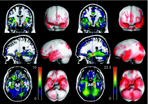
Methods: Voxel-based morphometry analyses of magnetic resonance images of the brain were performed to determine the correlation between regional extra-hippocampal grey matter volume and hippocampal grey matter volume. The results from 36 patients with right and left MTLE were compared, and results from the two groups were compared with those from 49 healthy controls.
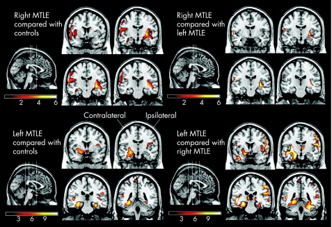
Results: Compared with controls, patients with MTLE showed a more intense correlation between hippocampal grey matter volume and regional grey matter volume in locations such as the contralateral hippocampus, bilateral parahippocampal gyri and frontal and parietal areas. Compared with right MTLE, patients with left MTLE exhibited a wider area of atrophy related to hippocampal grey matter loss, encompassing both the contralateral and ipsilateral hemispheres, particularly affecting the contralateral hippocampus.
Conclusions: Our results suggest that left hippocampal atrophy is associated with a larger degree of extra-hippocampal atrophy. This may help to explain the more intense cognitive impairment usually observed in these patients.
Conflict of interest statement
Competing interests: None declared.
Similar articles
-
An optimized voxel-based morphometric study of gray matter changes in patients with left-sided and right-sided mesial temporal lobe epilepsy and hippocampal sclerosis (MTLE/HS).Epilepsia. 2010 Apr;51(4):511-8. doi: 10.1111/j.1528-1167.2009.02324.x. Epub 2009 Oct 8. Epilepsia. 2010. PMID: 19817822
-
Distribution of regional gray matter abnormalities in a pediatric population with temporal lobe epilepsy and correlation with neuropsychological performance.Epilepsy Behav. 2007 Dec;11(4):558-66. doi: 10.1016/j.yebeh.2007.07.005. Epub 2007 Oct 22. Epilepsy Behav. 2007. PMID: 17933587
-
Voxel-based morphometry reveals gray matter network atrophy in refractory medial temporal lobe epilepsy.Arch Neurol. 2004 Sep;61(9):1379-84. doi: 10.1001/archneur.61.9.1379. Arch Neurol. 2004. PMID: 15364683
-
Meta-analysis of voxel-based morphometry studies of gray matter abnormalities in patients with mesial temporal lobe epilepsy and unilateral hippocampal sclerosis.Brain Imaging Behav. 2018 Oct;12(5):1497-1503. doi: 10.1007/s11682-017-9797-5. Brain Imaging Behav. 2018. PMID: 29302917
-
MTLE with hippocampal sclerosis in adult as a syndrome.Rev Neurol (Paris). 2015 Mar;171(3):259-66. doi: 10.1016/j.neurol.2015.02.004. Epub 2015 Feb 26. Rev Neurol (Paris). 2015. PMID: 25727907 Review.
Cited by
-
Temporal lobe epilepsy affects spatial organization of entorhinal cortex connectivity.Epilepsy Behav. 2018 Nov;88:87-95. doi: 10.1016/j.yebeh.2018.06.038. Epub 2018 Sep 20. Epilepsy Behav. 2018. PMID: 30243111 Free PMC article.
-
Graph theory findings in the pathophysiology of temporal lobe epilepsy.Clin Neurophysiol. 2014 Jul;125(7):1295-305. doi: 10.1016/j.clinph.2014.04.004. Epub 2014 Apr 21. Clin Neurophysiol. 2014. PMID: 24831083 Free PMC article. Review.
-
Remote effects of hippocampal damage on default network connectivity in the human brain.J Neurol. 2009 Dec;256(12):2021-9. doi: 10.1007/s00415-009-5233-0. Epub 2009 Jul 15. J Neurol. 2009. PMID: 19603243
-
Relationship Between Seizure Frequency and Functional Abnormalities in Limbic Network of Medial Temporal Lobe Epilepsy.Front Neurol. 2019 May 8;10:488. doi: 10.3389/fneur.2019.00488. eCollection 2019. Front Neurol. 2019. PMID: 31133978 Free PMC article.
-
Reduced Interhemispheric White Matter Asymmetries in Medial Temporal Lobe Epilepsy With Hippocampal Sclerosis.Front Neurol. 2019 Apr 24;10:394. doi: 10.3389/fneur.2019.00394. eCollection 2019. Front Neurol. 2019. PMID: 31068889 Free PMC article.
References
-
- Meencke H J V G. Hippocampal sclerosis in epilepsy. In: Luders HO, ed. Epilepsy surgery. New York: Raven Press, 1991705–715.
MeSH terms
LinkOut - more resources
Full Text Sources