. 2006;46(2):113-22.
doi: 10.1385/CBB:46:2:113.

Chronotropic response of cultured neonatal rat ventricular myocytes to short-term fluid shear

Affiliations

Chronotropic response of cultured neonatal rat ventricular myocytes to short-term fluid shear

Ilka Lorenzen-Schmidt et al. Cell Biochem Biophys. 2006.

Abstract

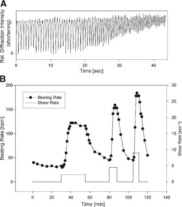
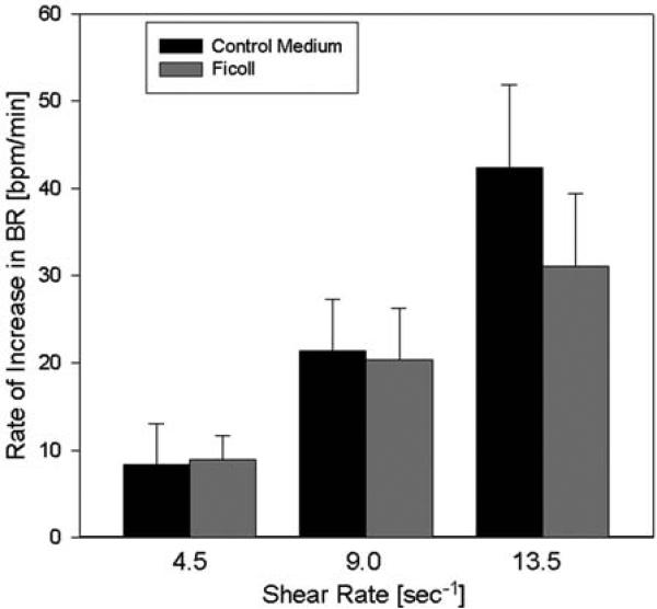
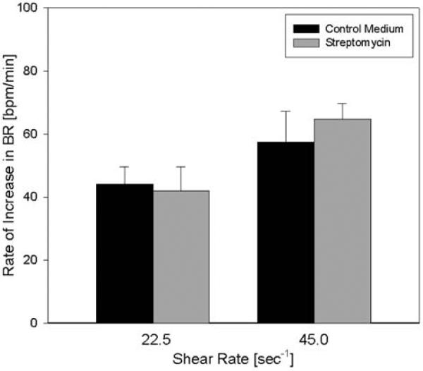
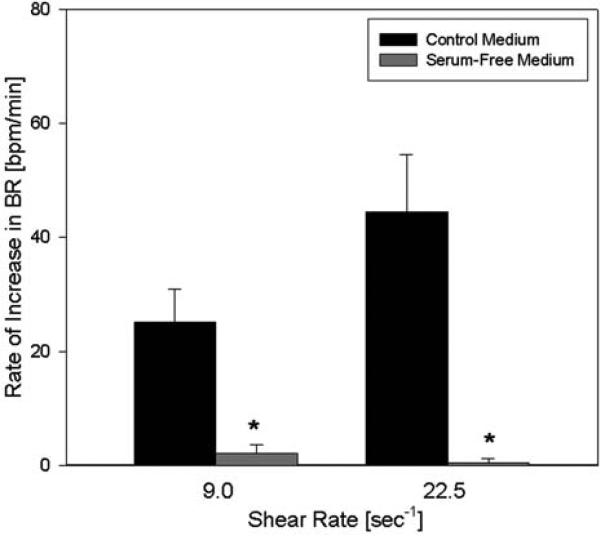
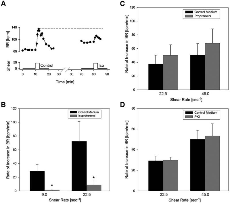
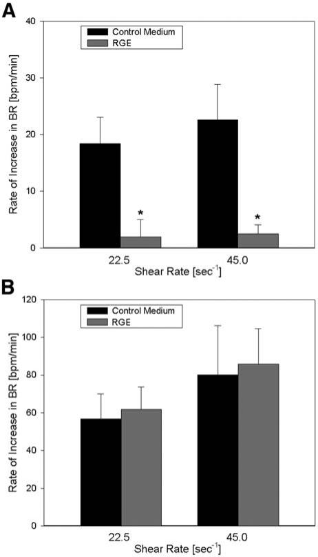
Ventricular myocytes are continuously exposed to fluid shear in vivo by relative movement of laminar sheets and adjacent cells. Preliminary observations have shown that neonatal myocytes respond to fluid shear by increasing their beating rate, which could have an arrhythmogenic effect under elevated shear conditions. The objective of this study is to investigate the characteristics of the fluid shear response in cultured myocytes and to study selected potential mechanisms. Cultured neonatal rat ventricular myocytes that were spontaneously beating were subjected to low shear rates (5-50/s) in a fluid flow chamber using standard culture medium. The beating rate was measured from digital microscopic recordings. The myocytes reacted to low shear rates by a graded and reversible increase in their spontaneous beating rate of up to 500%. The response to shear was substantially attenuated in the presence of the beta-adrenergic agonist isoproterenol (by 86+/-8%), as well as after incubation with integrin-blocking RGD peptides (by 92+/-8%). The results suggest that the beta-adrenergic signaling pathway and integrin activation, which are known to interact, may play an important role in the response mechanism.


Figures

Fig. 1
Fig. 1
(A) Shortening of a representative group of cultured neonatal rat cardiomyocytes, showing the increase in contraction frequency during shear exposure (shear rate: 45/s, shear stress: 450 mdyn/cm2). Because shortening was measured as relative light diffraction intensity from randomly oriented myocytes, the unit is arbitrary. As the beating rate increased, the myocytes became unable to relax completely, leading to a diminished amplitude of shortening. (B) Representative changes in spontaneous beating rate from different shear rates (2.25, 4.5, and 9/s) in cultured ventricular myocytes. The response is consistent, reversible, and graded by the magnitude of shear.
Fig. 2
Fig. 2
Effect of viscosity changes on the shear response. The viscosity was increased 2.5-fold by addition of the macromolecule ficoll (0.1 nM), resulting in higher shear stress at the same shear rate. For instance, at a shear rate of 9.0/s, shear stress amounted to 90 mdyn/cm2 in control medium and to about 225 mdyn/cm2 with added Ficoll. The rate of shear response was not significantly affected by the increase in viscosity (n = 6).
Fig. 3
Fig. 3
Effect of streptomycin on the shear response. Myocytes were incubated with the stretch-activated channel blocker streptomycin (70 μM) and exposed to shear. Streptomycin did not significantly affect the rate of response to shear at either shear rate compared with control medium (n = 3).
Fig. 4
Fig. 4
Effect of serum-free medium on the response to shear. Myocytes were exposed to shear with control medium and serum-free medium. The increase in beating rate with shear was nearly abolished at both shear rates in the absence of serum compared with the control medium (n = 4). Asterisks denote statistical significance from control (p < 0.05).
Fig. 5
Fig. 5
Impact of isoproterenol, propranolol and PKI on the response to shear. (A) Example of the contraction rate response to shear in the absence and presence of isoproterenol (iso). Note that the increase with isoproterenol does not reach the maximum beating rate. (B) Summarized response to isoproterenol: after incubation with isoproterenol (1 μM), the response to shear was significantly blunted compared with control medium at both shear rates (n = 6). Asterisks denote statistical significance from control medium (p < 0.05). (C) Shear response in control medium compared with medium containing the nonspecific β-blocker propranolol (1 μM). Propranolol (n = 5) did not affect the rate of response to shear significantly. (D) Effect of PKA-inhibiting peptide PKI (5 μM) on the response. No statistical difference was detected (n = 4).
Fig. 6
Fig. 6
(A) Effect of RGD peptide on the rate of response to shear. β1-integrin-blocking RGD peptide (100 μM) nearly abolished the shear response (n = 5) compared with control medium at both shear rates. The asterisk denotes a statistical difference (p < 0.05) from control. (B) Effect of RGE peptide on the response to shear. The non-integrin-binding RGE peptide (100 μM) was used as an additional control (n = 5) and did not have a significant effect on the beating rate increase at either shear rate.

References

    1. Dou J, Tseng WY, Reese TG, Wedeen VJ. Combined diffusion and strain MRI reveals structure and function of human myocardial laminar sheets in vivo. Magn. Reson. Med. 2003;50:107–113. - PubMed
    1. Dewey CF, Jr., Bussolari SR, Gimbrone MA, Jr., Davies PF. The dynamic response of vascular endothelial cells to fluid shear stress. J. Biomech. Eng. 1981;103:177–185. - PubMed
    1. Sterpetti AV, Cucina A, D'Angelo LS, Cardillo B, Cavallaro A. Response of arterial smooth muscle cells to laminar flow. J. Cardiovasc. Surg. (Torino) 1992;33:619–624. - PubMed
    1. Moazzam F, DeLano FA, Zweifach BW, Schmid-Schonbein GW. The leukocyte response to fluid stress. Proc. Natl. Acad. Sci. USA. 1997;94:5338–5343. - PMC - PubMed
    1. Coughlin MF, Schmid-Schonbein GW. Pseudopod projection and cell spreading of passive leukocytes in response to fluid shear stress. Biophys. J. 2004;87:2035–2042. - PMC - PubMed

Publication types