. 2006 Nov;7(11):1209-16.
doi: 10.1038/ni1392. Epub 2006 Oct 1.

Apoptotic neutrophils and T cells sequester chemokines during immune response resolution through modulation of CCR5 expression

Affiliations

Apoptotic neutrophils and T cells sequester chemokines during immune response resolution through modulation of CCR5 expression

Amiram Ariel et al. Nat Immunol. 2006 Nov.

Abstract

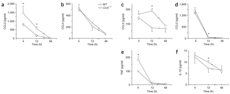
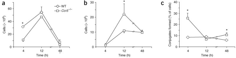
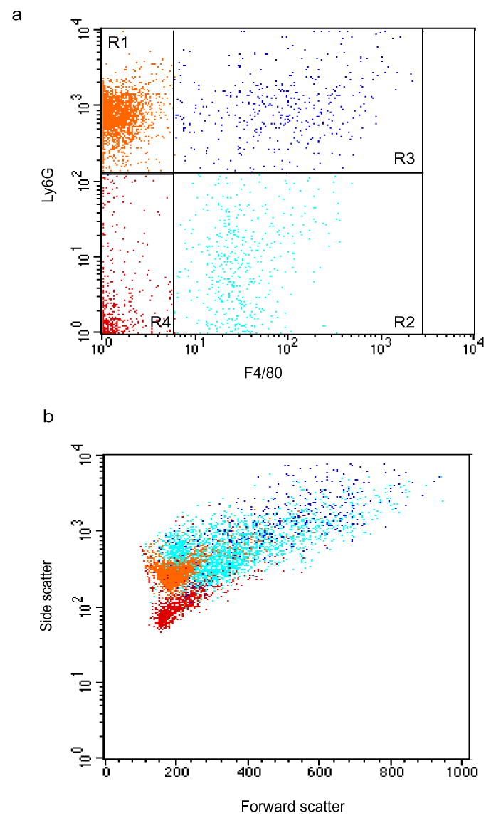
During the resolution phase of inflammation, the 'corpses' of apoptotic leukocytes are gradually cleared by macrophages. Here we report that during the resolution of peritonitis, the CCR5 chemokine receptor ligands CCL3 and CCL5 persisted in CCR5-deficient mice. CCR5 expression on apoptotic neutrophils and activated apoptotic T cells sequestered and effectively cleared CCL3 and CCL5 from sites of inflammation. CCR5 expression on late apoptotic human polymorphonuclear cells was downregulated by proinflammatory stimuli, including tumor necrosis factor, and was upregulated by 'proresolution' lipid mediators, including lipoxin A4, resolvin E1 and protectin D1. Our results suggest that CCR5+ apoptotic leukocytes act as 'terminators' of chemokine signaling during the resolution of inflammation.


Conflict of interest statement

COMPETING INTERESTS STATEMENT

The authors declare competing financial interests (see the Nature Immunology website for details).

Figures

Figure 1
Figure 1
Increased CCL3 and CCL5 in peritoneal exudates from Ccr5−/− mice during the resolution of peritonitis. Peritoneal exudates from Ccr5−/− or wild-type (WT) mice were collected 4, 12 and 48 h after the initiation of peritonitis and CCL3 (a), CCL4 (b), CCL5 (c), CCL2 (d), TNF (e) and IL-1β (f) were measured by Luminex methodology. *, P < 0.05, wild-type versus Ccr5−/−. Results are the mean ± s.e.m. of three experiments.
Figure 2
Figure 2
Ccr5−/− macrophages engulf apoptotic PMNs more efficiently than do wild-type macrophages. Flow cytometry of PMNs (a), monocytes and macrophages (b) and PMN-macrophage conjugates (c) from peritoneal exudates of Ccr5−/− or wild-type mice, stained with anti-Ly6G and anti-F4/80. *, P < 0.05, wild-type versus Ccr5−/−. Results are the mean ± s.e.m. of three experiments.
Figure 3
Figure 3
Ccr5−/− macrophages are more mature than wild-type macrophages. (a,b) Flow cytometry of cells from the peritoneal exudates of wild-type mice (a) and Ccr5−/− mice (b), stained with anti-Ly6G and anti-F4/80 and analyzed 48 h after initiation of peritonitis. Numbers for outlined areas indicate percent Ly6G+F4/80+ cells (top) or Ly6GF4/80+ cells (bottom). (c,d) Average mean fluorescence intensity (MFI) of F4/80 staining on monocytes and macrophages (c) and PMN-macrophage conjugates (d) at various time points (horizontal axes). *, P < 0.05, wild-type versus Ccr5−/−. Results are representative of three experiments (a,b) and the mean ± s.e.m. of three experiments (c,d).
Figure 4
Figure 4
CCR5 mediates chemokine scavenging by apoptotic PMNs.(a) Chemokines and cytokines in peritoneal exudates of recipients of PMNs sorted from exudates from Ccr5−/− or wild-type mice 4 h after peritonitis initiation and cultured overnight, then transferred into the peritonea of Ccr5−/− mice 12 h after the initiation of peritonitis; peritoneal exudates of recipient mice were collected 1 h later for analysis. *, P < 0.05, wild-type versus Ccr5−/− PMN recipients. (b) Chemokines and cytokines in peritoneal exudates of recipients of PMN-enriched exudate cells collected 12 h after peritonitis induction, treated with either CCR5 antagonist or negative control and then transferred into inflamed wild-type peritonea 4 h after injection of recipient mice with zymosan A; peritoneal exudates of recipient mice were collected 1 h later for analysis. *, P < 0.05, control versus CCR5 antagonist treatment. Results are mean ± s.e.m. of four experiments.
Figure 5
Figure 5
Regulation of CCR5 expression on late apoptotic human PMNs. (a,b) Flow cytometry of CCR5 surface expression (a) and apoptotic phenotype (b) of human PMNs incubated for 22 h with vehicle, zVAD-fmk or TNF. (c) Flow cytometry of CCR5 surface expression and apoptotic phenotype of human PMNs exposed for 22 h to vehicle, resolution-phase lipid mediators or TGF-β. RvE1-Me, resolvin E1–methyl ester; ATLa, aspirin-triggered lipoxin A4 analog; PD1-Me, PD1–methyl ester. *, P < 0.05, vehicle versus experimental treatment. Results are representative of three experiments (a,b) and the mean ± s.e.m. of three experiments (c).
Figure 6
Figure 6
Apoptotic activated peripheral blood T cells have high expression of CCR5. (a) Flow cytometry of the surface phenotype of peripheral blood T cells activated for 3 d by anti-CD3 (5 μg/ml). Gray bar, percent death of all cells. (b,c) Flow cytometry of CCR5 surface expression (b) and apoptotic surface phenotype (c) of T cells activated as described in a and treated for 4–48 h with staurosporine (STS). *, P < 0.05, versus late apoptotic. Results are representative of three experiments.
Figure 7
Figure 7
Late apoptotic T cells have high expression of CCR5. Flow cytometry of CCR5 surface expression and apoptotic phenotypes of Jurkat CD4+ T cells incubated with vehicle (0.2% DMSO for 48 h; ac), staurosporine (2 μM for 24 h; df) or Fas ligand (5 ng/ml for 48 h; g,h) and then stained with propidium iodide (ag) and mouse IgG (a,d,g), anti-CCR5 (b,e,h) or anti-CXCR4 (c,f). Positively stained late apoptotic cells are outlined. Results are representative of ten experiments.
Figure 8
Figure 8
CCR5 on apoptotic cells has characteristics different from those of CCR5 on live cells. (a) Flow cytometry of CCL4 binding and apoptotic phenotype of T cells incubated for 48 h with staurosporine or vehicle, then incubated with increasing concentrations of biotinylated CCL4 followed by fluorescein isothiocyanate–avidin. *, P < 0.05, versus live cells. (b) Light microscopy (left) and fluorescent microscopy (middle and right) of live cells (top) or apoptotic cells (bottom) from a that were fixed after staining and applied to coverslips. Arrowheads indicate CCR5 clustering. PI, propidium iodide. Original magnification, ×1,000. Results represent mean ± s.e.m. of three experiments (a) or are representative of three experiments (b).
Supplementary Figure 1
Supplementary Figure 1. Ly-6G+F4/80+ in peritoneal exudates are PMN-macrophage conjugates
Leukocytes recovered from peritoneal exudates 48 h after peritonitis initiation were stained with anti-Ly-6G and anti-F4/80 and analyzed by flow cytometry. Dot plots represent Ly-6G and F4/80 staining (a) and forward versus side scatter (b). PMNs (R1, orange cells), macrophages (R2, cyan cells) and PMN-macrophage conjugates (R3, blue cells) are indicated. Results are respresentative of six experiments.
Supplementary Figure 2
Supplementary Figure 2. Increased CCL4 binding to late apoptotic T cells
(a–c) Late apoptotic (a) or live and early apoptotic (b) Tc ells were incubated with STS, indicated concentrations of Fas ligand, or vehicle for 24 h. Cells were then incubated with biotinylated CCL4 or soybean trypsin inhibitor (STI) as a nonspecific biotinylated probe, followed by FITC-avidin. After washing, CCL4 binding (a,b) and apoptotic phenotype (c) was assessed by flow cytometry. *P < 0.05 versus vehicle alone. Results are representative of six experiments.
Supplementary Figure 3
Supplementary Figure 3. Caspase inhibition abrogates apoptosis-induced modulation of CCL4 binding to late apoptotic T cells
T cells were incubated with vehicle (a–d) or Z-VAD-fmk (e,f) for 20 min and then treated with vehicle (a,b), STS (24 h; c,e) or Fas ligand (48 h; d,f). Cells were incubated with biotinylated CCL4 (open histograms) or STI (filled histograms) and analyzed by flow cytometry. (g) Relative inhibiton of CCL4 binding and apoptosis induced by Z-VAD-fmk. *P < 0.05 versus CCL4 binding. Results are representative of (a–f) and present the mean ± s.e.m. of (g) three experiments.
Supplementary Figure 4
Scheme of the role of apoptotic leukocytes and pro-resolving lipid mediators in the resolution of inflammation.

References

    1. Majno G, Joris I. Cells, Tissues, and Disease: Principles of General Pathology. Oxford University Press: New York; 2004.
    1. Serhan CN. Lipoxins and novel aspirin-triggered 15-epi-lipoxins (ATL): a jungle of cell-cell interactions or a therapeutic opportunity? Prostaglandins. 1997;53:107–137. - PubMed
    1. Levy BD, Clish CB, Schmidt B, Gronert K, Serhan CN. Lipid mediator class switching during acute inflammation: signals in resolution. Nat Immunol. 2001;2:612–619. - PubMed
    1. Serhan CN. A search for endogenous mechanisms of anti-inflammation uncovers novel chemical mediators: missing links to resolution. Histochem Cell Biol. 2004;122:305–321. - PubMed
    1. Rossi AG, Haslett C. In: Proinflammatory and Antiinflammatory Peptides. Said SI, editor. Marcel Dekker; New York: 1998. pp. 9–24.

Publication types

MeSH terms

LinkOut - more resources