. 2006 Nov 15;299(2):517-28.
doi: 10.1016/j.ydbio.2006.09.003. Epub 2006 Sep 9.

N-cadherin is required for neural crest remodeling of the cardiac outflow tract

Affiliations

N-cadherin is required for neural crest remodeling of the cardiac outflow tract

Yang Luo et al. Dev Biol. .

Abstract

Cardiac neural crest cells undergo extensive cell rearrangements during the formation of the aorticopulmonary septum in the outflow tract. However, the morphogenetic mechanisms involved in this fundamental process remain poorly understood. To determine the function of the Ca2+-dependent cell adhesion molecule, N-cadherin, in murine neural crest, we applied the Cre/loxP system and created mouse embryos genetically mosaic for N-cadherin. Specifically, deletion of N-cadherin in neural crest cells led to embryonic lethality with distinct cardiovascular defects. Neural crest cell migration and homing to the cardiac outflow tract niche were unaffected by loss of N-cadherin. However, N-cadherin-deficient neural crest cells were unable to undergo the normal morphogenetic changes associated with outflow tract remodeling, resulting in persistent truncus arteriosus in the majority of mutant embryos. Other mutant embryos initiated aorticopulmonary septum formation; however, the neural crest cells were unable to elongate and align properly along the midline and remained rounded with limited contact with their neighbors. Interestingly, rotation of the outflow tract was incomplete in these mutants suggesting that alignment of the channels is dependent on N-cadherin-generated cytoskeletal forces. A second cardiac phenotype was observed where loss of N-cadherin in the epicardium led to disruption of heterotypic cell interactions between the epicardium and myocardium resulting in a thinned ventricular myocardium. Thus, we conclude that in addition to its role in myocardial cell adhesion, N-cadherin is required for neural crest cell rearrangements critical for patterning of the cardiac outflow tract and in the maintenance of epicardial-myocardial cell interactions.


Figures

Figure 1
Figure 1
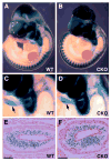
Loss of N-cadherin in NCCs leads to embryonic lethality. Whole-mount images of E10.5 (A), E11.5 (B) and E12.5 (C) wild-type (WT) and N-cadherin CKO (CKO) embryos. Note the pale appearance of the CKO compared to WT at E12.5.
Figure 2
Figure 2
Normal distribution pattern of NCC in E10.5 embryos after deletion of N-cadherin. Wnt1-Cre recombination in NCC is marked by the expression of β-galactosidase in wild-type (WT) and N-cadherin CKO (CKO) R26R embryos (A,B). Higher magnification of the branchial arches and outflow tract (arrow) in the X-gal stained embryos (C,D). Cross-section through the outflow tract of the X-gal stained embryos (E,F) indicating colonization by NCC. Scale bar: E,F 50 μm.
Figure 3
Figure 3
Deletion of N-cadherin in neural crest cells leads to truncus arteriosus. Histological analysis of serial frontal sections from WT (A,C,E) and N-cadherin CKO (B,D,F) E11.75 embryos. The OFT (arrow) is undivided in the CKO compared to the WT heart. Ao, ascending aorta; PA, pulmonary artery. Scale bar: 25 μm.
Figure 4
Figure 4
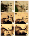
Undivided OFT in the N-cadherin/Wnt1-Cre mutant embryos. Left lateral view after intracardiac ink injection to visualize OFT and pharyngeal arch arteries at E10.5 (A,B) and E11.75 (C–F). The pharyngeal arch arteries are numbered. At E10.5, the developing OFT and pharyngeal arch arteries appeared similar in the WT and CKO embryos (n=2). The boxed region of the OFT (C,D) is shown at higher magnification (E,F). By E11.75, separation of the OFT (arrows) was observed in the WT embryo, in contrast, no apparent division of the OFT was seen in the mutant embryo (n=3). Ao, ascending aorta; PA, pulmonary artery.
Figure 5
Figure 5
Normal pattern of smooth muscle cells surrounding the pharyngeal arch arteries in N-cad/Wnt1-Cre embryos. Histological (A,C) and immunofluorescence (B,D) analysis was performed on adjacent frontal sections from E11.5 wild-type (A,B) and CKO (C,D) embryos (n=3). The pharyngeal arch arteries are numbered. The pattern of α-smooth muscle actin staining appeared similar in the wild-type (B) and CKO (D) embryos. Scale bar: 25 μm.
Figure 6
Figure 6
Abnormal outflow tract development in N-cadherin CKO embryos. Histological sections of outflow tract of wild-type (WT) and N-cadherin CKO (CKO) E12.5 embryos (A–D). Note the smaller, less developed outflow tract in the CKO compared to WT embryo. Higher magnification (inset) of the midline shows the rounded, less compacted morphology of the NCC in the CKO compared to WT embryo. Transmission electron microscopy of outflow tract (E,F) further illustrates the loss of cell-cell adhesion between the rounded CKO NCC compared to the elongated, aligned WT NCC. The differentiation marker, α-smooth muscle actin, was significantly reduced in the less compacted septum region of the CKO (H) compared to WT (G) embryo. Scale bars: A,B 25 μm; C,D 50 μm; E,F 2 μm; G,H 50 μm.
Figure 7
Figure 7
Cadherin/catenin expression in the NCC of the outflow tract. Immunofluorescence was performed on E10.5 (A,B), E11.5 (C,E,F), E12.5 (D) embryos for N-cadherin or β-catenin (E,F). N-cadherin was present at a low level in WT (arrow, inset) and absent from the NCC in the CKO (arrow, inset) embryo (A,B). N-cadherin expression dramatically increased at regions of cell-cell contact as the WT NCC rearranged in the outflow tract (C,D). Beta-catenin expression was significantly reduced in the N-cadherin-deficient NCC indicating it is the primary classical cadherin involved in outflow tract remodeling. Scale bars: 50 μm.
Figure 8
Figure 8
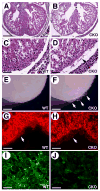
Loss of epicardial – myocardial cell interactions in the N-cadherin CKO embryos. Histological sections of wild-type (WT) and N-cadherin CKO (CKO) E12.5 hearts show a thinned myocardial wall associated with a detached epicardium (arrow) in the CKO (A–D). Note the incomplete development of the ventricular septum (asterik). Whole-mount images of the heart surface after removal of the pericardial cell layer illustrate the bubbling of the epicardium in the CKO (arrows) compared to the smooth surface of the WT heart (E,F). Immunofluorescence demonstrated that N-cadherin expression was lost in the epicardium (arrow) of the CKO embryo (H). Note the high level of N-cadherin in the underlying myocardium. To confirm the in vivo results, epicardial cells were isolated from hearts and immunostained for N-cadherin (I,J). Scale bars: A,B 12.5 μm; C,D,G,H,I,J 50 μm; E,F 20 μm.

References

    1. Akitaya T, Bronner-Fraser M. Expression of cell adhesion molecules during initiation and cessation of neural crest cell migration. Dev Dyn. 1992;194:12–20. - PubMed
    1. Borchers A, David R, Wedlich D. Xenopus cadherin-11 restrains cranial neural crest migration and influences neural crest specification. Development. 2001;128:3049–60. - PubMed
    1. Brault V, Moore R, Kutsch S, Ishibashi M, Rowitch DH, McMahon AP, Sommer L, Boussadia O, Kemler R. Inactivation of the beta-catenin gene by Wnt1-Cre-mediated deletion results in dramatic brain malformation and failure of craniofacial development. Development. 2001;128:1253–64. - PubMed
    1. Bronner-Fraser M, Wolf JJ, Murray BA. Effects of antibodies against N-cadherin and N-CAM on the cranial neural crest and neural tube. Dev Biol. 1992;153:291–301. - PubMed
    1. Chen TH, Chang TC, Kang JO, Choudhary B, Makita T, Tran CM, Burch JB, Eid H, Sucov HM. Epicardial induction of fetal cardiomyocyte proliferation via a retinoic acid-inducible trophic factor. Dev Biol. 2002;250:198–207. - PubMed

Publication types