

Contrast-enhanced ultrasound for examining tumor biology
- PMID: 17015239
- PMCID: PMC1693765
- DOI: 10.1102/1470-7330.2006.0023
Contrast-enhanced ultrasound for examining tumor biology
Abstract
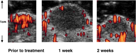
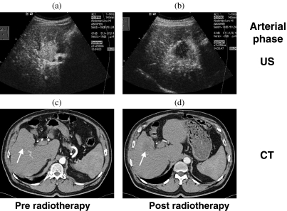
This paper reviews the potential of ultrasound for assessing the viability and biological behavior of tumors. Unlike color Doppler sonography, modern techniques for contrast-enhanced ultrasound permit the measurement of tissue perfusion irrespective of vessel size or flow velocity. Perfusion can also be assessed quantitatively, using replenishment kinetics or derivates thereof. The perfusion of tumors is a surrogate parameter of their viability and may mirror their response to therapy. Furthermore, the degree of vascularity in a tumor may express its aggressiveness and help to predict its response to treatment. In animal models, a decrease in blood flow has been shown to precede a shrinkage of tumors treated with anti-angiogenic compounds. In liver metastases, arterial and portal blood supply can be assessed separately, and a response to stereotactic radiotherapy was found to go along with a decrease in arterial perfusion. Moreover, a relatively high arterial perfusion of liver metastases may predict a response to chemotherapy. Contrast-enhanced ultrasound may be a potent tool for assessing the effects of anti-angiogenic treatment in patients.
(c) International Cancer Imaging Society.
Figures
References
-
- de Jong N, Frinking PJ, Bouakaz A, Goordern M, Schourmans T, Jingping X, et al. Optical imaging of contrast agent microbubbles in an ultrasound field with a 100-MHz camera. Ultrasound Med Biol. 2000;26:487–92. - PubMed
-
- Blomley M, Albrecht T, Cosgrove D, Jayaram V, Butler-Barnes J, Eckersley R. Stimulated acoustic emission in liver parenchyma with Levovist. Lancet. 1998;351:568. - PubMed
-
- Albrecht T, Blomley MJK, Heckemann RA, Cosgrove DO, Jayaram V, Patel N, et al. Stimulierte akustische Emission mit dem Ultraschallkontrastmittel Levovist: ein klinisch nutzbarer Effekt mit leberspezifischen Eigenschaften. Fortschr Röntgenstr. 2000;172:61–7. - PubMed
-
- Burns PN. Harmonic imaging with ultrasound contrast agents. Clin Radiol. 1996;51(1):50–55. - PubMed
-
- Hope Simpson D, Burns PN. Pulse inversion Doppler: a new method for detecting nonlinear echoes from microbubble contrast agents. IEEE Trans Ultrason Ferroelec Freq Contr. 1999;46:372–82. - PubMed
Publication types
MeSH terms
Substances
LinkOut - more resources
Full Text Sources