. 2006 Dec 22;3(11):805-21.
doi: 10.1098/rsif.2006.0144.

Bioactive and bioresorbable cellular cubic-composite scaffolds for use in bone reconstruction

Affiliations

Bioactive and bioresorbable cellular cubic-composite scaffolds for use in bone reconstruction

Yasuo Shikinami et al. J R Soc Interface. .

Abstract

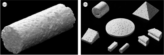
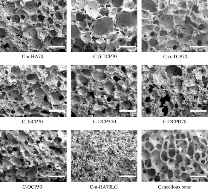
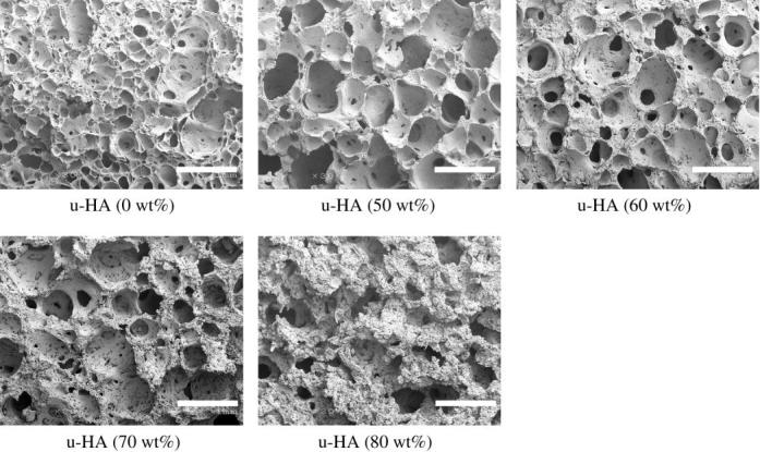
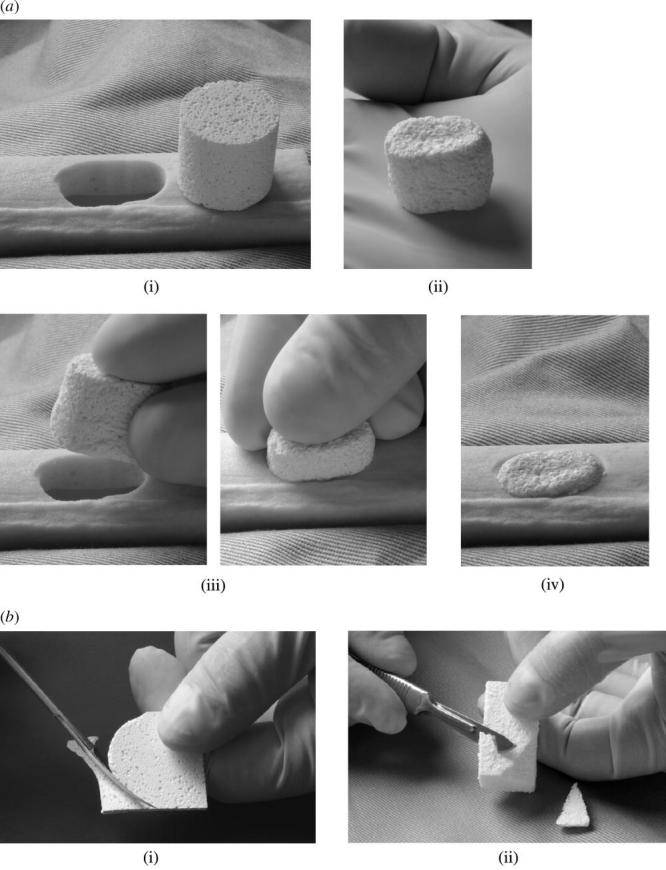
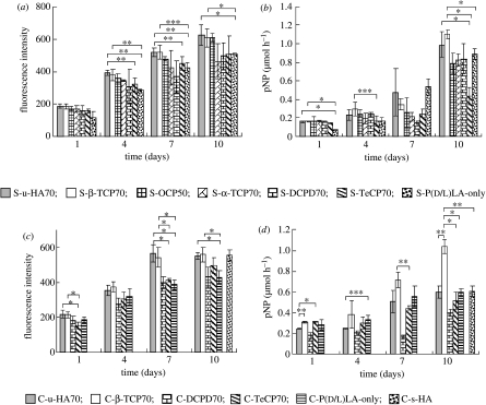
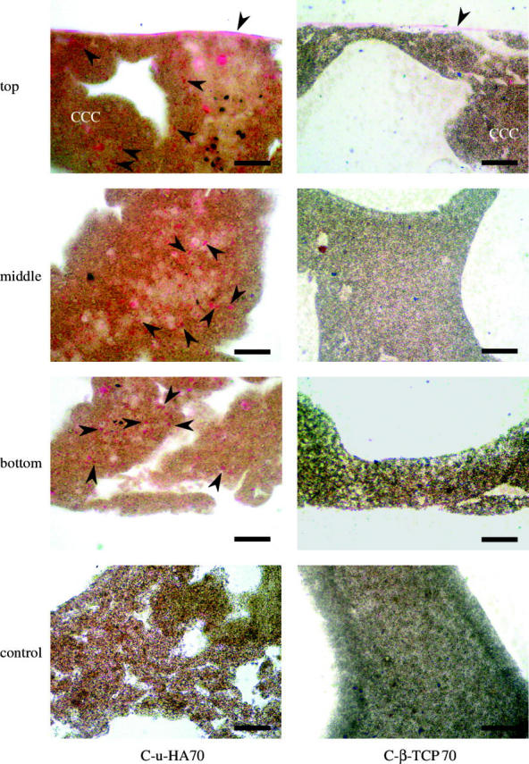
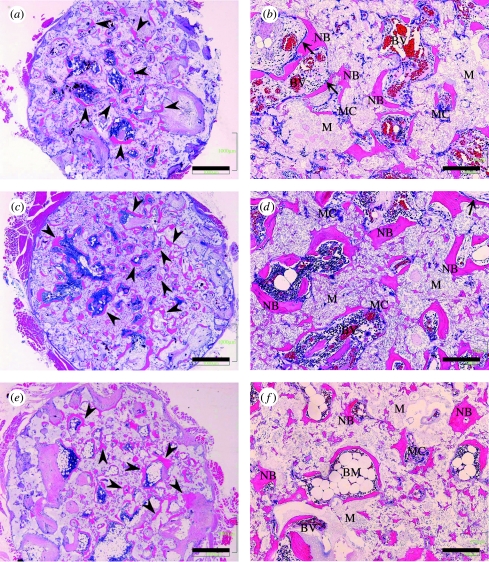
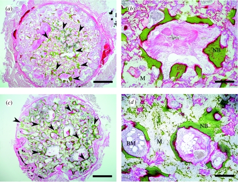
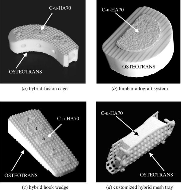
We used a novel composite fibre-precipitation method to create bioactive and bioresorbable cellular cubic composites containing calcium phosphate (CaP) particles (unsintered and uncalcined hydroxyapatite (u-HA), alpha-tricalcium phosphate, beta-tricalcium phosphate, tetracalcium phosphate, dicalcium phosphate dihydrate, dicalcium phosphate anhydrate or octacalcium phosphate) in a poly-D/L-lactide matrix. The CaP particles occupied greater than or equal to 70 wt% (greater than or equal to 50 vol%) fractions within the composites. The porosities of the cellular cubic composites were greater than or equal to 70% and interconnective pores accounted for greater than or equal to 70% of these values. In vitro changes in the cellular geometries and physical properties of the composites were evaluated over time. The Alamar Blue assay was used to measure osteoblast proliferation, while the alkaline phosphatase assay was used to measure osteoblast differentiation. Cellular cubic C-u-HA70, which contained 70 wt% u-HA particles in a 30 wt% poly-D/L-lactide matrix, showed the greatest three-dimensional cell affinity among the materials tested. This composite had similar compressive strength and cellular geometry to cancellous bone, could be modified intraoperatively (by trimming or heating) and was able to form cortico-cancellous bone-like hybrids. The osteoinductivity of C-u-HA70, independent of biological growth factors, was confirmed by implantation into the back muscles of beagles. Our results demonstrated that C-u-HA70 has the potential as a cell scaffold or temporary hard-tissue substitute for clinical use in bone reconstruction.


Figures

Figure 1
Figure 1
(a) Representative cylindrical block of C-β-TCP70. (b) Customized blocks of C-u-HA70 (Comporus).
Figure 2
Figure 2
SEM images of the inner pores of CaP particle/P(d/l)LA cellular cubic composites and cancellous bone of a beagle distal tibia (original magnification ×30; scale bar, 1 mm).
Figure 3
Figure 3
SEM images of inner pores of u-HA/P(d/l)LA cellular cubic composites with different u-HA contents (original magnification ×30; scale bar, 1 mm).
Figure 4
Figure 4
Pore-size distribution in C-u-HA70.
Figure 5
Figure 5
Changes in Mv¯ values of C-u-HA70, C-β-TCP70 and C-OCP50 over time in SBF at 37 °C.
Figure 6
Figure 6
Changes in Sc values of C-u-HA70, C-β-TCP70 and C-OCP50 over time in SBF at 37 °C. The symbols filled circle, filled square and filled triangle denote Sc, whereas open circle, open square and open triangle denote Sc retention.
Figure 7
Figure 7
In vitro cellular changes in C-u-HA70 under μCT with time (original magnification ×30; scale bar, 500 μm).
Figure 8
Figure 8
Intraoperative transformation and trimming procedures for C-u-HA70. (a) Transformation and insertion procedures: (i) a Comporus C-u-HA70 cylinder (ø18× H 18 mm) and a hole of rectangle with rounded ends in an artificial bone (minor axis =16 mm); (ii) the transformed oval column after heating at 65 °C; (iii) insertion of the column into the defect and (iv) the column set in the artificial bone. (b) Trimming procedures: (i) a square Comporus C-u-HA70 plate being trimmed into a round sheet using operating scissors; (ii) removal of the edges of a Comporus C-u-HA70 block by shaving with a scalpel.
Figure 9
Figure 9
Alamar Blue assay (fluorescence excitation wavelength, 544 nm; fluorescence emission wavelength, 590 nm) of HOBs in (a) sheet composites and (c) cellular cubic composites, and ALP activities of HOBs in (b) sheet composites and (d) cellular cubic composites, over 10 days. *p<0.005; **p<0.01; ***p<0.05.
Figure 10
Figure 10
Microscopic H&E-stained images of HOBs cultured on C-u-HA70 and C-β-TCP70 for 7 days. Arrowheads indicate HOBs (original magnification ×200; scale bar, 50 μm). HOBs were not seeded in the controls. As the organic solvent to get rid of the resin dissolves these cellular cubic composites, the sections were stained with H&E while embedded in resin. The HOBs therefore appeared to be lightly stained.
Figure 11
Figure 11
H&E staining of decalcified sections. Ectopic bone formation (arrow heads) was seen in the pores of C-u-HA70. Osteoblast-like cells (arrow) were detected adjacent to the newly formed bone. (a) and (b) Two months after implantation of C-u-HA70; (c) and (d) Three months after implantation of C-u-HA70; (e) and (f) Twelve months after implantation of C-u-HA70. (a), (c) and (e) Original magnification ×20; scale bar, 1 mm. (b), (d) and (f) Original magnification ×100; scale bar, 200 μm; BM, bone-marrow tissue; BV, blood vessel; M, material; MC, multinucleated cell; NB, newly formed bone.
Figure 12
Figure 12
Villanueva Goldner stain of undecalcified sections. Ectopic bone formation (arrow heads) was seen in the pores of C-u-HA70. (a) and (b) Three months after implantation of C-u-HA70; (c) and (d) Twelve months after implantation of C-u-HA70. (a) and (c) Original magnification ×20; scale bar, 1 mm. (b) and (d) Original magnification ×100; scale bar, 200 μm. BM, bone-marrow tissue; M, material; NB, newly formed bone.
Figure 13
Figure 13
Hybrid-type bone substitutes and scaffolds. Combinations of C-u-HA70 (Comporus) and OSTEOTRANS (u-HA/PLLA; 40 : 60 wt ratio).

References

    1. Ahmed S.A, Gogal R.M, Jr, Walsh J.E. A new rapid and simple non-radioactive assay to monitor and determine the proliferation of lymphocytes: an alternative to [3H]thymidine incorporation assay. J. Immunol. Methods. 1994;170:211–224. doi:10.1016/0022-1759(94)90396-4 - DOI - PubMed
    1. Bessey O.A, Lowry O.H, Brock M.J. A method for the rapid determination of alkaline phosphatase with five cubic millimeters of serum. J. Biol. Chem. 1946;164:321–329. - PubMed
    1. Burdick J.A, Frankel D, Dernell W.S, Anseth K.S. An initial investigation of photocurable three-dimensional lactic acid based scaffolds in a critical-sized cranial defect. Biomaterials. 2003;24:1613–1620. doi:10.1016/S0142-9612(02)00538-0 - DOI - PubMed
    1. Chang B.S, Lee C.K, Hong K.S, Youn H.J, Ryu H.S, Chung S.S, Park K.W. Osteoconduction at porous hydroxyapatite with various pore configurations. Biomaterials. 2000;21:1291–1298. doi:10.1016/S0142-9612(00)00030-2 - DOI - PubMed
    1. Eggli P.S, Muller W, Schenk R.K. Porous hydroxyapatite and tricalcium phosphate cylinders with two different pore size ranges implanted in the cancellous bone of rabbits. A comparative histomorphometric and histologic study of bony ingrowth and implant substitution. Clin. Orthop. Rel. Res. 1988;232:127–138. - PubMed

LinkOut - more resources