Review
. 2006 Dec 22;3(11):741-52.
doi: 10.1098/rsif.2006.0141.

Polypyrrole-based conducting polymers and interactions with biological tissues

Affiliations
Review

Polypyrrole-based conducting polymers and interactions with biological tissues

D D Ateh et al. J R Soc Interface. .

Abstract

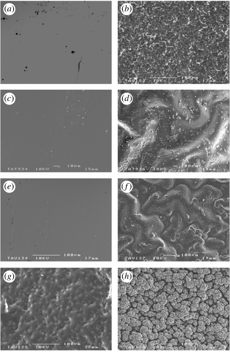
Polypyrrole (PPy) is a conjugated polymer that displays particular electronic properties including conductivity. In biomedical applications, it is usually electrochemically generated with the incorporation of any anionic species including also negatively charged biological macromolecules such as proteins and polysaccharides to give composite materials. In biomedical research, it has mainly been assessed for its role as a reporting interface in biosensors. However, there is an increasing literature on the application of PPy as a potentially electrically addressable tissue/cell support substrate. Here, we review studies that have considered such PPy based conducting polymers in direct contact with biological tissues and conclude that due to its versatile functional properties, it could contribute to a new generation of biomaterials.


Figures

Figure 1
Figure 1
Examples of conducting polymer structures. The conjugated structure consisting of an alternating carbon–carbon double bond is common to all conducting polymers.
Figure 2
Figure 2
Electropolymerizaion mechanism of polypyrrole. Monomer units are adsorbed onto the surface of the working electrode resulting in one-electron oxidation to form a pyrrole cation radical. These cations then couple with themselves, with other cations or with neutral monomers from solution. In each case, this leads to the formation of a dimer dication, which undergoes a double deprotonation to give a neutral molecule. These more stable dimer radicals have a lower oxidation potential compared with the monomer units and chain growth then occurs by preferential coupling between the dimers and monomers (Skotheim 1986). Anion (A) is required to maintain electroneutrality.
Figure 3
Figure 3
Scanning electron microscopy images of polypyrrole (PPy) surface topography generated for different counterions and electropolymerization durations (a–b) PPy/chloride, (c–d) PPy/polyvinyl sulphate, (e–f) PPy/dermatan and (g–h) PPy/collagen. The shorter times produced thin films (left column) with none or little surface features whereas at extended times, thicker films with distinct topography are seen (right column). More instances of counterion controlled topography may be found in the literature (Skotheim 1986).
Figure 4
Figure 4
In vivo tissue response to polypyrrole-hyaluronic acid (PPy/HA) bilayer films. (a) Polypyrrole-polystyrene sulphonate (PPy/PSS) films and (b) PPy/HA bilayer films were implanted into subcutaneous pouches in rats. Tissue surrounding the material was harvested after two weeks, fixed, imbedded and stained with hematoxylin and eosin. The heavy black lines in both images are the PPy/PSS and PPy/HA bilayer films. Blood vessels are denoted by arrows. Scale bar, 100 μm (both images are at the same magnification). This figure shows that HA retains its angiogenesis properties whilst incorporated in PPy since more blood vessels were seen around this implant compared to the PPy/PSS control. Reprinted from Collier et al. (2000) with permission from John Wiley & Sons, Inc.
Figure 5
Figure 5
Polypyrrole (PPy) implants. (a) An example of a typical implant; scale bar, 1 mm (b) two PPy implants placed in the rat's cortex; scale bar, 2 mm (c) a histological slice at six weeks post-implantation with the remnants of the Polypyrrole implant; scale bar, 200 μm. Favourable responses for PPy compared to Teflon implants in terms of macrophage activity, gliosis and neuronal integration were found after implantation. Reprinted from George et al. (2005) with permission from Elsevier.
Figure 6
Figure 6
Neuronal cell line adhesion and proliferation on different substrates after 8, 24 and 72 h of culture. The numbers of cells were normalized to initial density of seeded cells (200 000 cells ml−1) (n=3 per substrates). The volume of the cell suspension used is 100 μl. All results are given at ±5%. PEI is polyethyleneimine, PPy is polypyrrole, PPI is polypropyleneimine and FTO is fluorine-doped tin oxide. This study illustrates that under some conditions poor interactions may occur between polypyrrole and cells compared to other materials. Reprinted from Lakard et al. (2004) with permission from Elsevier.
Figure 7
Figure 7
(Left) Scanning electron microscopy of a PC-12 cell on polypyrrole (PPy). PC-12 cells were cultured in NGF-supplemented medium for 48 h on thick disks of PPy, then processed for scanning electron microscopy. Bar =10 μm. (Right) Neurite length histograms. Shown are histograms of neurite lengths for cells on PPy (a) with electrical stimulation (S) and (b) without (NS) potential applied through the PPy film. Histograms for cells on PPy with (c) potential applied through the solution and on (d) tissue culture polystyrene (TCPS) are also shown. This study demonstrates the potential of electrically stimulating PPy in order to affect cell behaviour. In this case, enhanced neurite outgrowth has implications in nerve regeneration therapies. Reprinted from Schmidt et al. (1997) with permission from National Academy of Sciences, USA (Copyright 1997).
Figure 8
Figure 8
Schematic of the cloud-like conducting polymer on the gold electrode polymerized through the hydrogel matrix. The flexibility and usefulness of polypyrrole may be further enhanced with such composites. Reprinted from Kim et al. (2004) with permission from John Wiley & Sons, Inc.

Similar articles

Cited by

References

    1. Adejolu S.B, Wallace G.G. Conducting polymers and the bioanalytical sciences: new tools for biomolecular communications—a review. Analyst. 1996;121:699–703. doi:10.1039/an9962100699 - DOI - PubMed
    1. Aoki T, Tanino M, Sanui K, Ogata N, Kumakura K, Okano T, Sakurai Y, Watanabe M. Culture of mammalian cells on polypyrrole-coated IT0 as a biocompatible electrode. Synth. Met. 1995;71:2229–2230. doi:10.1016/0379-6779(94)03235-X - DOI
    1. Aoki T, Tanino M, Sanui K, Ogata N, Kumakura K. Secretory function of adrenal chromaffin cells cultured on polypyrrole films. Biomaterials. 1996;17:1971–1974. doi:10.1016/0142-9612(96)00015-4 - DOI - PubMed
    1. Armes S.P. Optimum reaction conditions for the polymerization of pyrrole by iron (III) chloride in aqueous solution. Synth. Mat. 1987;20:365–371.
    1. Ateh D.D, Vadgama P, Navsaria H. Culture of human keratinocytes on polypyrrole-based conducting polymers. Tissue Eng. 2006;12:645–655. doi:10.1089/ten.2006.12.645 - DOI - PubMed

Publication types

LinkOut - more resources