. 2006 Oct 15;20(20):2806-19.
doi: 10.1101/gad.1459706. Epub 2006 Oct 2.

The bHLH-PAS protein Spineless is necessary for the diversification of dendrite morphology of Drosophila dendritic arborization neurons

Affiliations

The bHLH-PAS protein Spineless is necessary for the diversification of dendrite morphology of Drosophila dendritic arborization neurons

Michael D Kim et al. Genes Dev. .

Abstract

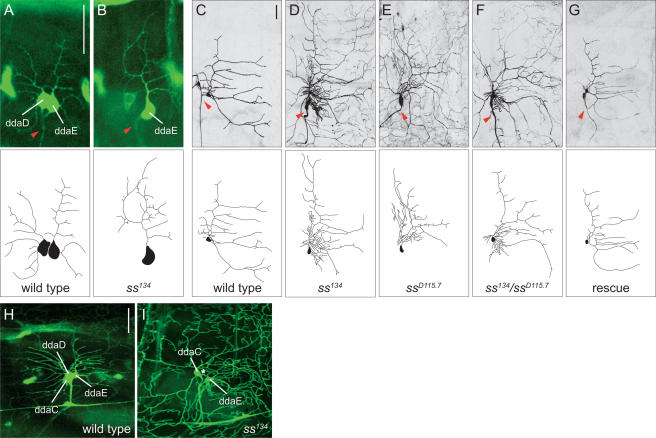
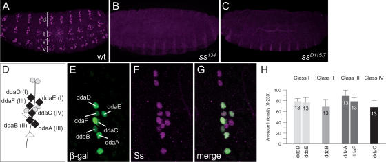
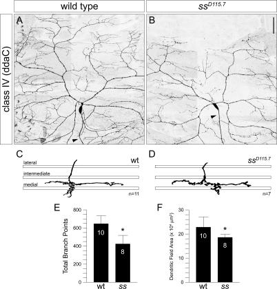
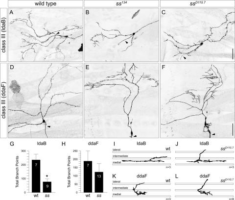
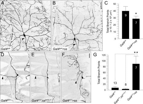
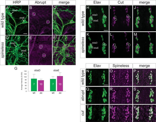
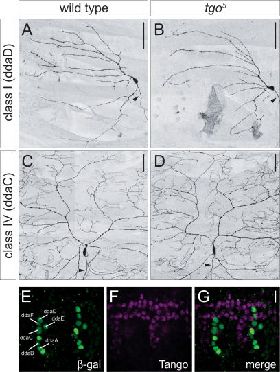
Dendrites exhibit a wide range of morphological diversity, and their arborization patterns are critical determinants of proper neural connectivity. How different neurons acquire their distinct dendritic branching patterns during development is not well understood. Here we report that Spineless (Ss), the Drosophila homolog of the mammalian aryl hydrocarbon (dioxin) receptor (Ahr), regulates dendrite diversity in the dendritic arborization (da) sensory neurons. In loss-of-function ss mutants, class I and II da neurons, which are normally characterized by their simple dendrite morphologies, elaborate more complex arbors, whereas the normally complex class III and IV da neurons develop simpler dendritic arbors. Consequently, different classes of da neurons elaborate dendrites with similar morphologies. In its control of dendritic diversity among da neurons, ss likely acts independently of its known cofactor tango and through a regulatory program distinct from those involving cut and abrupt. These findings suggest that one evolutionarily conserved role for Ahr in neuronal development concerns the diversification of dendrite morphology.


Figures

Figure 1.
Figure 1.
spineless regulates the dendritic morphogenesis of class I da neurons. (A) Wild-type dorsal class I neurons ddaD and ddaE in a stage 17 (18–20 h AEL) embryo visualized by the Gal4221 reporter. (B) ddaE neuron in a homozygous ss134 mutant exhibits overgrowth of the primary branch and increased terminal branching. (C) Wild-type ddaE neuron in a third instar larva. (D–F) ddaE neurons in a homozygous ss134 mutant (D), ssD115.7 mutant (E), and ss134/ssD115.7 transheterozygous mutant (F) exhibit overgrowth of the primary branch and increased terminal branching. (G) Expressing UAS-ss with Gal4221 in ss mutants rescues the primary branch overgrowth and increased terminal branching phenotype. Red arrowheads indicate axons. (H) Expression of GFP in the dorsal class I neurons (ddaD and ddaE) in a second instar larva as visualized by the Gal4221 reporter. GFP is weakly expressed in the dorsal class IV neuron ddaC at this developmental stage. (I) Expression of GFP in a homozygous ss134 mutant as visualized by the Gal4221 reporter. GFP is highly expressed in ddaE and ddaC, while absent in ddaD (normal position is indicated by a white asterisk). The terminal branches of ddaC are clearly visible. Anterior is to the left, and dorsal is up in this and all subsequent figures. Bar, 25 μm.
Figure 2.
Figure 2.
Spineless expression in the embryonic PNS. (A) Expression of Ss protein in stage 16 (16–18 h AEL) embryo. Ss is expressed in all sensory neurons in the dorsal (d), lateral (l), ventral prime (v′), and ventral (v) clusters. (B,C) Ss expression is absent in homozygous ss134 (B) and ssD115.7 (C) mutants. (D) Schematic depiction of sensory neurons in the dorsal cluster. Black diamonds represent da neurons, gray circles represent es organs, and white triangles represent bd neurons. (E) All multidendritic (md) neurons in the dorsal cluster in a stage 16 embryo labeled by the E7-2-36 lacZ enhancer trap line as revealed by staining with β-gal. (F) Expression of Ss in the dorsal cluster. (G) Merged image of E and F showing expression of Ss in the da neurons. (H) Quantification of Spineless immunoreactivity (measured as mean pixel intensity) in dorsal da neurons.
Figure 3.
Figure 3.
spineless functions cell-autonomously to regulate dendritic branching in class I and II da neurons. (A) Normal morphology of class I neuron ddaE dendrites in a wild-type third instar larva MARCM clone. ddaE normally projects its secondary dendrites toward the posterior segment border and elaborates few terminal branches. (B,C) ddaE neurons exhibit increased terminal branching in ss134 (B) and ssD115.7 (C) MARCM clones. (D) Quantification of the total number of branchpoints for ddaE in wild-type and ssD115.7 clones. Error bars represent standard deviation in this and all subsequent figures (asterisk; p < 0.001, two- tailed t-test). (E) Normal morphology of class I neuron ddaD dendrites in a wild-type MARCM clone. ddaD normally projects its secondary dendrites toward the anterior segment border. (F,G) ddaD neurons show increased branching and dendrite routing defects in ss134 (F) and ssD115.7 (G) MARCM clones. Secondary dendrites are misoriented, and branches improperly terminate posterior to the cell body. (H) Quantification of total number of branchpoints for ddaD in wild- type and ssD115.7 clones (asterisk; p < 0.001, two-tailed t-test). (I) Normal morphology of dorsal class II neuron ddaB dendrites in a wild-type MARCM clone. ddaB extends long primary and secondary dendrites with few terminal branches. (J,K) ddaB neurons exhibit increased dendritic branching at the distal tips of primary and secondary dendrites in ss134 (J) and ssD115.7 (K) MARCM clones. (L) Quantification of total number of branchpoints for ddaB in wild-type and ssD115.7 clones (asterisk; p < 0.001, two-tailed t-test). Arrowheads indicate axons. Bar, 50 μm.
Figure 4.
Figure 4.
Loss of spineless function reduces the complexity of class IV dendrites. (A) Normal morphology of class IV neuron ddaC dendrites in a wild-type MARCM clone. ddaC extends its dendrites to the dorsal midline and anterior and posterior segment borders. (B) A ddaC ssD115.7 MARCM clone shows reduced terminal branching and failure of dendrites to reach the dorsal midline. Arrowheads indicate axons. (C,D) Tracing of representative ddaC axon terminals in wild-type (C) and ssD115.7 (D) MARCM clones. The three bars represent the lateral, intermediate, and medial fascicles of the VNC as revealed by FasII immunostaining. ddaC axon terminals in wild-type (C) and ssD115.7 (D) MARCM clones invariably terminate near the medial fascicle. (E) Quantification of total number of branchpoints for ddaC in wild-type and ssD115.7 clones (asterisk; p < 0.001, two- tailed t-test). (F) Quantification of total dendritic field area for ddaC in wild-type and ssD115.7 clones (asterisk; p < 0.02, two- tailed t-test). Bar, 50 μm.
Figure 5.
Figure 5.
spineless regulates the number of dendritic spikes in the class III neurons. (A) Normal morphology of lateral class III neuron ldaB dendrites in a wild-type MARCM clone. Dendritic spikes are found along all major branches. (B,C) ldaB neurons show a reduction of dendritic spikes along all major branches in ss134 (B) and ssD115.7 (C) MARCM clones. (D) Normal morphology of dorsal class III neuron ddaF dendrites in a wild-type MARCM clone. ddaF extends long primary and secondary dendrites that are decorated with dendritic spikes. (E,F) ddaF neurons show a reduction in the number of dendritic spikes and exhibit clusters of longer terminal branches along the primary dendrites in ss134 (E) and ssD115.7 (F) MARCM clones. Arrowheads indicate axons. (G) Quantification of total number of branchpoints for ldaB in wild-type and ssD115.7 clones (asterisk; p < 0.001, two-tailed t-test). (H) Quantification of total number of branchpoints for ddaF indicates no significant difference between wild-type and ssD115.7 clones. (I,J) Tracing of representative ldaB axon terminals in wild-type (I) and ssD115.7 (J) MARCM clones. ldaB axons always terminate between the intermediate and medial fascicles in wild-type (I) and ssD115.7 (J) MARCM clones. (K,L) Tracing of representative ddaF axon terminals in wild-type (K) and ssD115.7 (L) MARCM clones. ddaF axons project into the VNC and curve posteriorly between the intermediate and medial fascicles in wild-type (K) and ssD115.7 (L) MARCM clones. Bar, 50 μm.
Figure 6.
Figure 6.
Overexpression of Spineless leads to opposite phenotypes in different sensory neurons. (A) Wild- type morphology of the class IV neuron ddaC as revealed by the Gal4ppk reporter line. (B) Overexpression of Ss in ddaC causes a severe reduction in terminal branching. (C) Quantification of total number of branchpoints for ddaC in wild type (Gal4ppk) and over-expressing Ss (Gal4ppk>ss) (asterisk; p < 0.001, two-tailed t-test). (D) Wild-type morphology of the td neuron as revealed by the Gal4477 reporter line. (E) The td neuron does not show any dendrite defects in ssD115.7 mutants. (F) Overexpression of Ss in the td neuron causes ectopic dendritic branches to form. (G) Quantification of total number of branchpoints for the td neuron in wild-type (Gal4477), ssD115.7 mutant (Gal4477; ssD115.7), and overexpressing Ss (Gal4477>ss) (asterisks; p < 0.001, two tailed t-test). Arrowheads indicate axons. Bar, 50 μm.
Figure 7.
Figure 7.
spineless acts through a regulatory program distinct from cut and abrupt to control dendritic diversity. (A) Wild-type third instar larva stained with the pan-neuronal marker horseradish peroxidase (HRP) indicating positions of class I neurons in dorsal cluster (ddaD and ddaE). (B) Wild-type expression pattern of Ab in the dorsal cluster. (C) Merged image of A and B showing that Ab is only expressed in the class I neurons. (D) HRP stain of homozygous ssD115.7 mutant third instar larva indicating the positions of ddaD and ddaE. (E) Expression pattern of Ab in the dorsal cluster in a homozygous ssD115.7 mutant. (F) Merged image of D and E showing Ab expression in the dorsal class I neurons in the ssD115.7 mutant. (G) Quantification of Ab immunoreactivity in class I neurons ddaD and ddaE in wild type and ss mutants reveals similar levels of Ab expression. (H) Wild-type embryo (18–20 h AEL) stained with the pan-neuronal marker Elav. (I) Wild-type expression pattern of Cut in the dorsal cluster. Cut is not expressed in the class I neurons ddaD and ddaE (circles indicate positions). (J) Merged image of H and I showing different levels of Cut expression in the da neurons. (K) Homozygous ssD115.7 mutant embryo stained with Elav. (L) Expression pattern of Cut in the dorsal cluster in a homozygous ssD115.7 mutant. Cut is not expressed in ddaD and ddaE (circles). (M) Merged image of K and L showing that Cut expression is normal in the ssD115.7 mutant. (N) Wild-type expression pattern of Elav in the dorsal cluster. (O) Wild-type expression pattern of Ss. (P) Merged image of N and O showing Ss expression in all Elav-positive neurons. (Q) Expression pattern of Elav in abK02807 loss-of-function mutant. (R) Expression pattern of Ss in the abK02807 mutant. (S) Merged image of Q and R showing that Ss expression is not regulated by ab in the sensory neurons. (T) Expression pattern of Elav in cutc145 loss-of-function mutant. (U) Expression pattern of Ss in the cutc145 mutant. (V) Merged image of T and U showing that Ss expression is not regulated by cut in the sensory neurons. Bar, 10 μm.
Figure 8.
Figure 8.
tgo does not act cell-autonomously to regulate the development of da neuron dendrites. (A) Wild-type MARCM clone of the class I neuron ddaD. (B) The ddaD tgo5 MARCM clone does not exhibit any dendrite branching or routing defects. (C) Wild-type MARCM clone of the class IV neuron ddaC. (D) The ddaC tgo5 MARCM clone does not show a reduction in terminal branching or dendritic field size. Arrowheads indicate axons. Bar, 50 μm. (E) Multidendritic (md) neurons in the dorsal cluster labeled by the E7- 2-36 lacZ enhancer trap line as revealed by staining with β-gal. (F) Wild-type expression pattern of Tgo. Tgo is largely accumulated in tracheal cells. (G) Merged image of E and F showing that Tgo is not accumulated in the nuclei of da neurons. Bar, 10 μm.

Comment in

References

    1. Ainsley, J.A., Pettus, J.M., Bosenko, D., Gerstein, C.E., Zinkevich, N., Anderson, M.G., Adams, C.M., Welsh, M.J., Johnson, W.A. Enhanced locomotion caused by loss of the Drosophila DEG/ENaC protein Pickpocket1. Curr. Biol. 2003;13:1557–1563. - PubMed
    1. Bodmer, R., Jan, Y.N. Morphological differentiation of the embryonic peripheral neurons in. Drosophila. Rouxs Arch.Dev. Biol. 1987;196:69–77. - PubMed
    1. Brenman, J.E., Gao, F.B., Jan, L.Y., Jan, Y.N. Sequoia, a tramtrack-related zinc finger protein, functions as a panneural regulator for dendrite and axon morphogenesis in. Drosophila. Dev. Cell. 2001;1:667–677. - PubMed
    1. Bridges, C.B., Morgan, T.H. Carnegie Institute of Washington; Washington DC: 1923. The third-chromosome group of mutant characters of Drosophila melanogaster; p. Publ. No. 327.
    1. Brown, R.P., McDonnell, C.M., Berenbaum, M.R., Schuler, M.A. Regulation of an insect cytochrome P450 monooxygenase gene (CYP6B1) by aryl hydrocarbon and xanthotoxin response cascades. Gene. 2005;358:39–52. - PubMed

Publication types