. 2006 Oct 1;20(19):2687-700.
doi: 10.1101/gad.1447006.

A positive feedback loop between the p53 and Lats2 tumor suppressors prevents tetraploidization

Affiliations

A positive feedback loop between the p53 and Lats2 tumor suppressors prevents tetraploidization

Yael Aylon et al. Genes Dev. .

Abstract

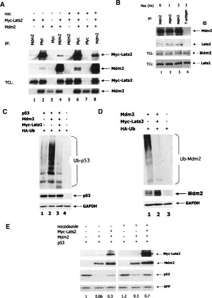
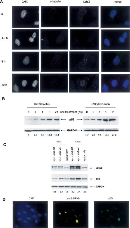
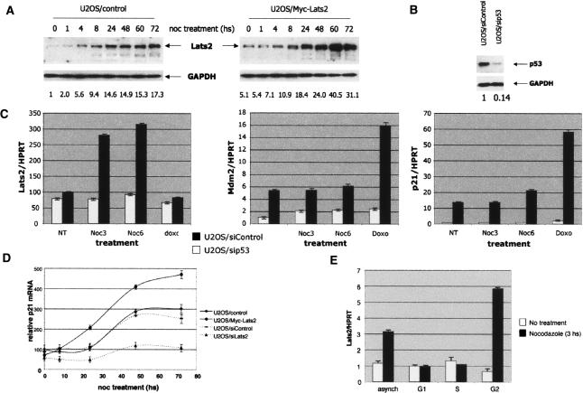
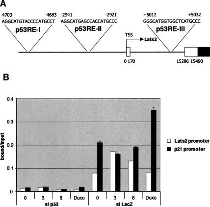
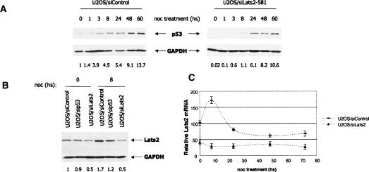
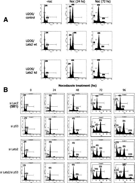
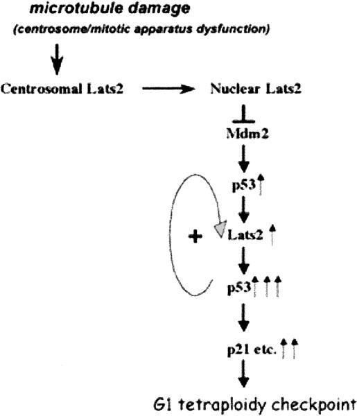
Damage to the mitotic spindle and centrosome dysfunction can lead to cancer. To prevent this, cells trigger a succession of checkpoint responses, where an initial mitotic delay is followed by slippage without cytokinesis, spawning tetraploid G1 cells that undergo a p53-dependent G1/S arrest. We describe the importance of Lats2 (Large Tumor Suppressor 2) in this checkpoint response. Lats2 binds Mdm2, inhibits its E3 ligase activity, and activates p53. Nocodazole, a microtubule poison that provokes centrosome/mitotic apparatus dysfunction, induces Lats2 translocation from centrosomes to the nucleus and p53 accumulation. In turn, p53 rapidly and selectively up-regulates Lats2 expression in G2/M cells, thereby defining a positive feedback loop. Abrogation of Lats2 promotes accumulation of polyploid cells upon exposure to nocodazole, which can be prevented by direct activation of p53. The Lats2-Mdm2-p53 axis thus constitutes a novel checkpoint pathway critical for the maintenance of proper chromosome number.


Figures

Figure 1.
Figure 1.
Lats2 binds Mdm2, inhibits its E3 ligase activity, and stabilizes p53. (A) Transfected Mdm2 and Lats2 interact in cells. HEK293 cells were transiently transfected with the indicated combinations of expression plasmids encoding Myc epitope-tagged human Lats2 (Myc-Lats2) and Mdm2. Twenty-four hours later, cells were treated with 0.2 μg/mL nocodazole for an additional 24 h, or mock-treated. Cell lysates were immunoprecipitated either with a mix of 4B2 and 4B11 anti-Mdm2 monoclonal antibodies (MoAbs) or with 9E10 anti-Myc MoAb. Immunoprecipitated proteins were resolved by SDS-PAGE and subjected to Western blot analysis, along with 2.5% of each total cell lysate (TCL). Mdm2 was detected with a mix of the anti-Mdm2 MoAbs 4B2 and 4B11; Myc-Lats2 was detected using the 9E10 MoAb. (B) Endogenous Mdm2 and Lats2 interact within cells. Endogenous Mdm2 was precipitated from U2OS cell extracts using a mix of 4B2, 4B11, and 2A9 MoAbs. Immunoprecipitation with MoAb against T-antigen (PAb419) was used as a negative control. Immunoprecipitated proteins and 3% of each TCL were detected using antibodies against Mdm2 (4B2, 4B11, and 2A9) or Lats2 (LA-2). (C) Lats2 inhibits p53 ubiquitination. p53/mdm2 double-null MEFs were transiently transfected with the indicated combinations of plasmids encoding p53, Mdm2, Myc-Lats2, and HA-tagged ubiquitin (400 ng, 200 ng, 8 μg, and 1.5 μg, respectively). Forty-eight hours later, cells were harvested. Extracts were denatured and immunoprecipitated with antibodies against p53 (mix of DO1 and PAb1801). Ubiquitinated forms of precipitated p53 were visualized using an anti-HA polyclonal Ab. Western blot of TCL was carried out as described in B. (D) Lats2 inhibits Mdm2 ubiquitination. p53/mdm2 double-null MEFs were transiently transfected with the indicated combinations of plasmids encoding Mdm2, MycLats2, and HA-tagged ubiquitin (400 ng, 8 μg, and 1.5 μg, respectively). Forty-eight hours later, cells were harvested, and extracts were denatured and immunoprecipitated with antibodies against Mdm2 (mix of 4B2, 4B11, and 2A9). Ubiquitinated forms of precipitated Mdm2 were visualized using an anti-HA polyclonal Ab. Western blot of TCL was carried out as described in B. (E) Lats2 overexpression increases p53 levels. p53/mdm2 double-null MEFs were transiently transfected with GFP (100 ng) expression plasmid together with the indicated combinations of p53, Mdm2, and Myc-Lats2 expression plasmids (40 ng, 40 ng, and 5 μg, respectively). Twenty-four hours later, cells were treated with nocodazole (0.2 μg/mL) or mock-treated for 20 h, harvested, and subjected to Western blot analysis. Quantification of relative p53 levels is shown below each lane.
Figure 2.
Figure 2.
Nocodazole induces Lats2 nuclear translocation and accumulation of Lats2 and p53. (A) Nocodazole induces Lats2 nuclear translocation. U2OS cells stably transfected with Myc-tagged Lats2 (U2OS/Myc-Lats2) were seeded on cover-slips. Twenty-four hours later, nocodazole (0.2 μg/mL) was added, and cells were fixed at the indicated times thereafter and subjected to immunofluorescence staining. Centrosomes were visualized with anti-γ-tubulin antibodies; Myc-Lats2 was visualized with anti-KPM (Santa Cruz Bio-technology) antibodies; nuclei were visualized with DAPI. Representative fields are shown (magnification 100×). The arrow indicates a centrosome negative for Lats2 staining. (B) Kinetics of p53 induction by nocodazole. U2OS/Myc-Lats2 cells and control cells (U2OS stably transfected with pcDNA3 empty vector) were treated with 0.2 μg/mL nocodazole (noc) and harvested at the indicated times. Endogenous p53 was detected with a mix of DO1 and PAb1801 MoAbs; endogenous GAPDH was used as a loading control (Chemicon). Quantification of relative p53 levels is shown below each lane. (C) Kinase activity of Lats2 is essential for p53 induction by nocodazole. U2OS cells were transiently transfected with wild-type (wt) or kinase-dead (KD) Lats2 or vector-only control. Twenty-four hours after transfection, transfected cells were selected for 3 d with 1 μg/mL puromycin. On the second day of puromycin treatment, nocodazole (0.2 μg/ mL) was added for 24 h. Endogenous p53 was visualized with DO1 and PAb1801 MoAbs; endogenous GAPDH was used as a loading control. Quantification of relative p53 levels is shown below each lane. (D) Nuclear accumulation of Lats2 correlates with p53 induction. U2OS cells were exposed to nocodazole, fixed after 8 h (magnification 40×), and analyzed as in A. Endogenous p53 and Lats2 were visualized with DO1 + PAb1801 or anti-KPM antibodies, respectively. A representative field is shown.
Figure 3.
Figure 3.
Rapid transcriptional activation of Lats2 is p53 dependent, specific for nocodazole, and occurs preferentially at the G2/M phase of the cell cycle. (A) Kinetics of Lats2 induction by nocodazole. U2OS/Myc-Lats2 cells and control cells were treated and analyzed as in Figure 2B. Lats2 (endogenous as well as stably transfected Myc-Lats2) was detected with LA-2 anti-Lats2 antibodies; endogenous GAPDH was used as a loading control. Quantification of relative Lats2 levels is shown below each lane. (B) U2OS cells were infected with a recombinant retrovirus encoding p53-specific shRNA (U2OS/sip53) or shRNA against lacZ (U2OS/siControl). From 24 h after infection, cells were maintained under puromycin selection. Endogenous p53 levels were monitored by Western blot analysis using DO1 + PAb1801; endogenous GAPDH was used as a loading control. Quantification of relative p53 levels is shown below each lane. (C) lats2 mRNA levels increase in a p53-dependent manner upon nocodazole treatment. U2OS cells with shRNA against lacZ (U2OS/siControl) and their p53-depleted derivatives (U2OS/sip53) were treated with 0.2 μg/mL nocodazole (noc) and harvested after 3 h (Noc3) or 6 h (Noc6) of treatment or exposed to 1 μM doxorubicin (Doxo) for 6 h. RNA was extracted and subjected to quantitative real-time RT–PCR, using primers specific for lats2, mdm2, and p21 mRNA. All values were normalized for the amount of HPRT RNA in the same sample. (D) Basal Lats2 levels modulate the extent of p21 mRNA induction by nocodazole. Stably transfected U2OS/control (transfected with pcDNA3), U2OS/Myc-Lats2, U2OS/siControl (shRNA against mouse PUMA, nonreactive with human PUMA mRNA), or U2OS/siLats2 (shRNA against lats2) cells were treated with 0.2 μg/mL nocodazole and harvested at the indicated times. RNA was extracted from each sample and subjected to quantitative real-time RT–PCR analysis, using primers specific for p21 mRNA. Values were normalized for the amount of HPRT RNA in the same sample. “relative p21 mRNA” represents normalized RNA levels compared with t = 0 levels in U2OS/control cells. (E) Lats2 transcription is induced by nocodazole solely in cells positioned in the G2/M phases of the cell cycle. U2OS cells were fractionated by centrifugal elutriation (see Supplementary Fig. 6). Mock-elutriated asynchronous cells (asynch), or G1-, S-, or G2/M-enriched populations were harvested after 3 h of treatment with 0.2 μg/mL nocodazole. RNA was extracted from each sample and subjected to quantitative real-time RT–PCR analysis, using primers specific for lats2 mRNA. Values were normalized for the amount of HPRT RNA in the same sample.
Figure 4.
Figure 4.
p53 associates with the Lats2 promoter in vivo. (A) Location and sequence of predicted p53REs in the Lats2 gene. Numbers represent nucleotide positions relative to the accepted 5′ end of the Lats2 mRNA transcription start site (TSS). Open boxes represent untranslated regions, and dark boxes represent coding regions. (B) ChIP analysis of DNA derived from stably transfected U2OS/sip53 or U2OS/siControl (siLacZ) cells. Cells were treated with 0.2 μg/mL nocodazole (noc) for 3 or 6 h or with 1 μM doxorubicin (Doxo) for 6 h. p53-bound chromatin was precipitated using CM1 polyclonal antibody (Novocastra). Chromatin-derived DNA was subjected to real-time PCR analysis with primer pairs bracketing the 5′ p53RE of the p21 gene and the Lats2 p53RE-I (see A). Values represent the ratio of the value obtained for bound DNA over that obtained for total input (chromatin before immunoprecipitation) DNA for each PCR primer pair (bound/input).
Figure 5.
Figure 5.
Lats2 is required for optimal induction of p53, p21, and Lats2 itself in response to nocodazole. (A) Optimal induction of p53 protein by nocodazole is compromised by Lats2 knockdown. Stably transfected U2OS/siControl and U2OS/siLats2-581 cells were treated with 0.2 μg/mL nocodazole (noc) and harvested at the indicated times. Endogenous p53 was detected using DO1 + PAb18O1 antibodies, and GAPDH was used as a loading control. Quantification of relative p53 levels is shown below each lane. (B) Full induction of Lats2 by nocodazole is dependent on p53 and Lats2 itself. Stably transfected U2OS cells subjected to the indicated shRNA knockdown were treated with 0.2 μg/mL nocodazole (noc) and harvested after 8 h. Endogenous Lats2 was visualized using LA-2 antibodies, and GAPDH was used as a loading control. (C) Basal Lats2 is required for efficient induction of subsequent Lats2 mRNA by nocodazole (positive feedback). Stably transfected U2OS/siControl and U2OS/siLats2 cells were treated with 0.2 μg/mL nocodazole (noc) and harvested at the indicated times. RNA was analyzed by quantitative RT–PCR, as in Figure 3D. “Relative Lats2 mRNA” represents HPRT-normalized RNA levels compared with t = 0 levels in siControl cells.
Figure 6.
Figure 6.
Lats2 modulates polyploidy and apoptosis upon prolonged treatment with nocodazole. (A) Lats2 overexpression prevents polyploidy and apoptosis in U2OS cells in response to nocodazole treatment. U2OS cells stably transfected with vector only (control), wild-type Myc-Lats2 (Lats2-wt), or kinase-dead Myc-Lats2 (Lats2-kd) were treated with 0.2 μg/mL nocodazole (noc) and harvested at the indicated times. Fixed cells were stained with propidium iodide (PI) to measure DNA content and subjected to FACS analysis. Horizontal bars mark areas quantified to determine the proportion of cells with the indicated DNA content. Asterisks indicate apoptotic cells with sub-G1 DNA content. (B) Cells lacking Lats2 reduplicate their DNA similarly to p53-null cells. Stably transfected U2OS/siControl (with shRNA against LacZ), U2OS/sip53, U2OS/siLats2 (using siLats2-581), and U2OS/siLats2/sip53 cells were treated with 0.2 μg/mL nocodazole (noc) and harvested at the indicated times. Fixed cells were stained with propidium iodide (PI) to measure DNA content and subjected to FACS analysis.
Figure 7.
Figure 7.
Model for the proposed roles of Lats2 and p53 in the response to microtubule-damage-induced stress. In response to microtubule damage and ensuing centrosome/mitotic apparatus dysfunction, Lats translocates from the centrosomes to the nucleus, where it binds Mdm2 and inactivates it. This leads to p53 activation, which selectively turns on Lats2 transcription, resulting in general up-regulation of nuclear Lats2 levels and further stabilization and induction of p53. The potent p53 activation leads to transcription of many other p53 target genes, including p21, and consequent arrest of tetraploid cells at the G1/S boundary, practically constituting a “G1 tetraploidy checkpoint.” Upward arrows indicate activation; the number of arrows indicates the relative extent of the activation. The positive feedback loop between p53 and Lats2 is also indicated.

References

    1. Abe, Y., Ohsugi, M., Haraguchi, K., Fujimoto, J., Yamamoto, T. LATS2–Ajuba complex regulates γ-tubulin recruitment to centrosomes and spindle organization during mitosis. FEBS Lett. 2006;580:782–788. - PubMed
    1. Andreassen, P.R., Lohez, O.D., Margolis, R.L. G2 and spindle assembly checkpoint adaptation, and tetraploidy arrest: Implications for intrinsic and chemically induced genomic instability. Mutat. Res. 2003;532:245–253. - PubMed
    1. Aronheim, A. Membrane recruitment systems for analysis of protein–protein interactions. Methods Mol. Biol. 2001;177:319–328. - PubMed
    1. Barr, F.A., Sillje, H.H., Nigg, E.A. Polo-like kinases and the orchestration of cell division. Nat. Rev. Mol. Cell Biol. 2004;5:429–440. - PubMed
    1. Bond, G.L., Hu, W., Levine, A.J. MDM2 is a central node in the p53 pathway: 12 years and counting. Curr. Cancer Drug Targets. 2005;5:3–8. - PubMed

Publication types

MeSH terms