. 2006 Oct 1;20(19):2739-53.
doi: 10.1101/gad.1444706.

Sustained Notch signaling in progenitors is required for sequential emergence of distinct cell lineages during organogenesis

Affiliations

Sustained Notch signaling in progenitors is required for sequential emergence of distinct cell lineages during organogenesis

Xiaoyan Zhu et al. Genes Dev. .

Abstract

Mammalian organogenesis results from the concerted actions of signaling pathways in progenitor cells that induce a hierarchy of regulated transcription factors critical for organ and cell type determination. Here we demonstrate that sustained Notch activity is required for the temporal maintenance of specific cohorts of proliferating progenitors, which underlies the ability to specify late-arising cell lineages during pituitary organogenesis. Conditional deletion of Rbp-J, which encodes the major mediator of the Notch pathway, leads to premature differentiation of progenitor cells, a phenotype recapitulated by loss of the basic helix-loop-helix (bHLH) factor Hes1, as well as a conversion of the late (Pit1) lineage into the early (corticotrope) lineage. Notch signaling is required for maintaining expression of the tissue-specific paired-like homeodomain transcription factor, Prop1, which is required for generation of the Pit1 lineage. Attenuation of Notch signaling is necessary for terminal differentiation in post-mitotic Pit1+ cells, and the Notch-repressed Pit1 target gene, Math3, is specifically required for maturation and proliferation of the GH-producing somatotrope. Thus, sustained Notch signaling in progenitor cells is required to prevent conversion of the late-arising cell lineages to early-born cell lineages, permitting specification of diverse cell types, a strategy likely to be widely used in mammalian organogenesis.


Figures

FIGURE 1
FIGURE 1
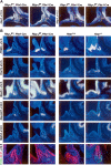
In situ analysis of expression pattern of core components of the Notch signaling pathway during pituitary development. Two ligands (Dll1, Jag1), two receptors (Notch2, Notch3), and the downstream target of Notch/Rbp-J signaling (Hes1) are expressed in the oral ectoderm and the Rathke's pouch (RP) by E12.5. At E13.5, Dll1, Jag1, Notch2, Notch3, and Hes1 expression are down-regulated in the anterior pituitary (AP) and are further confined to the perilumenal cells. At E14.5, Jag1 is expressed in the mesenchymal cells surrounding and within the pituitary gland. (IL) Intermediate lobe.
FIGURE 2
FIGURE 2
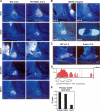
Notch signaling represses premature corticotrope differentiation via its downstream target Hes1. (A) Hes1 and Hey1 are down-regulated in Rbp-J f/f, Pitx1-Cre mice at E11.5. (B) Premature corticotrope differentiation is indicated by POMC as well as Tbx19 expression in Rbp-J f/f, Pitx1-Cre mice at E12.5. Expression of bHLH genes, including Mash1, NeuroD1, and Math3, are up-regulated but restricted in the ventral region of Rathke's pouch. Corticotrope premature differentiation is evident in Hes1 - / - mice, accompanied by up-regulation of Tbx19, Mash1, and NeuroD1. However, there is no pronounced ectopic expression of Math3. The posterior lobe of pituitary of Hes1 - / - mice is absent. Double-immunofluorescence staining of POMC and Ki67 in E12.5 embryos showed increased POMC in Rbp-J f/f, Pitx1-Cre and Hes1 - / - embryos in comparison with their respective littermate controls. POMC+ cells are Ki67-. Cells surrounding the lumen remain proliferative, while more cells at the ventral region of the pouch in mutant embryos exit the cell cycle and are negative for Ki67 staining.
FIGURE 3
FIGURE 3
Notch signaling is required for Pit1 lineage commitment. (A) Pit1 expression is absent in Rbp-J f/f, Pitx1-Cre mutant embryos at E13.5. At E17.5, Rbp-J f/f, Pitx1-Cre mutant embryos, Pit1 expression, and differentiation of Pit1 lineages thyrotropes (TSHβ) and somatotropes (GH) are impaired, while gonadotrope commitment, indicated by SF1 expression at E14.5 and LH expression at E17.5, is not affected in Rbp-J f/f, Pitx1-Cre mutant embryos. (B) Pit1 induction occurs normally in E13.5 Hes1 -/- embryos. (C) Double-immunofluorescence staining of POMC (green) and Ki67 (red), or Pit1 (green) and Ki67 (red) at E14.5 pituitaries shows that the anterior pituitary of the Rbp-J f/f, Pitx1-Cre mutant is populated with an increased number of corticotropes and is devoid of Pit1+ cells. The differentiated cells are not proliferative, as indicated by Ki67 staining. Cells surrounding the lumen are Ki67+ in both Rbp-J f/f, Pitx-Cre mutant embryos and littermate controls. (D) D2R expression at the intermediate lobe (IL) is increased in Rbp-J f/f, Pitx1-Cre mutant embryos at E17.5. Dual-immunofluorescence labeling of POMC (green) and Ki67 (red) at E17.5 pituitaries shows almost all POMC+ cells in IL are Ki67-.
FIGURE 4
FIGURE 4
Prop1 is a direct target of Notch signaling. (A) Prop1 expression is significantly down-regulated in Rbp-J f/f, Pitx1-Cre mutant embryos at E12.5 but is unchanged at E11.5. (B) Axin2 expression in Rbp-J f/f, Pitx1-Cre mutant embryos and Prop1 expression in Hes1 -/- at E12.5 are not significantly changed. (C) Genomic DNA sequences of mouse and human Prop1 were compared using VISTA. (Red) Promoters, (yellow) UTRs, (blue) exons, (pink) introns. Two evolutionarily conserved regions, the promoter and the first intron, show >75% homology. A putative Rbp-J-binding site is identified in the first intron. (D) A 32P-labeled 25-bp oligonucleotide encompassing the putative Rbp-J-binding site was incubated in the absence (lane 1) or in the presence (lanes 2-5) of in vitro translated Rbp-J and the competitors. Unlabeled oligonucleotides (lane 3), equivalent oligonucleotides where the putative Rbp-J-binding site was mutated (lane 4), or the oligonucleotides containing a Rbp-J-binding site from Epstein-Barr virus C promoter region (lane 5) were used as competitors at 100× molar excess. The arrow indicates the shifted probe caused by Rbp-J binding, and the arrowhead indicates free probe. (E) Quantitative ChIP assay of E12.5 pituitaries using anti-Rbp-J showed recruitment of Rbp-J to the first intron of Prop1. The promoter regions of the Hes1 and GHRHR were used as the positive and negative controls, respectively. (F,G) Transient transfection of 2-kb Prop1-Luc (F) or Prop1in-Luc (G) with control siRNA or siRNA specific to Rbp-J.
FIGURE 5
FIGURE 5
(A) Transient transfection in the pituitary GC cell line shows that the Pit1-NICD transgene construct can stimulate Hes1-Luc activity. Immunohistochemical staining with anti-HA antibody to detect transgene expression in E17.5 transgenic pituitary. Transgene expression is detectable as early as E13.5 when Pit1 expression begins. (B) Double-immunofluorescence staining of Pit1 (green) and Ki-67 (red) in wild-type and transgenic pituitary at E14.5 and E17.5 showed almost all proliferating cells are Pit1-negative at these stages and ectopic NICD expression in Pit1 lineage did not induce proliferation. (C) Prolonged activation of Notch signaling inhibits terminal cell differentiation of Pit1 lineages. In situ analysis of the transgenic pituitaries was performed with the GH, TSHβ, POMC, LHβ, and GHRHR. Differentiation of corticotropes and gonadotropes is less affected. (D) Double-immunofluorescence staining of HA (red) and terminal differentiation markers GH (left, green) and TSHβ (right, green) in pituitaries of transgenic mice showed that differentiated cells did not express the transgene.
FIGURE 6
FIGURE 6
(A) In situ analysis of bHLHs expression in E14.5 pituitaries of transgenic mice and wild-type control. Both Hes1 and Hes5 were induced by ectopic NICD expression while Mash1, NeuroD1, and Math3 were significantly repressed. (B) Math3 expression ontogeny during pituitary development. Expression of Math3 begins at E13.5 in the caudomedial region of the anterior pituitary and persists in the anterior lobe of the adult pituitary. (C) Expression of Math3 is down-regulated in Snell mice at E17.5. (D) Alignment of the promoter regions of human and mouse Math3 using VISTA identified an evolutionarily conserved region in the promoter with >75% homology. A putative binding site for Pit1 lies in this region. (E) ChIP from adult pituitaries using anti-Pit1 shows Pit1 is recruited to the Math3 as well as GHRHR promoter regions.
FIGURE 7
FIGURE 7
(A) Math3 is required for somatotrope maturation. In situ analysis in E17.5 pituitaries of Math3 -/- and littermate controls showed that somatotrope markers GH and GHRHR are down-regulated in Math3 -/-. Immunofluorescence staining showed GH protein is undetectable at E17.5 in Math3 -/- embryos. GHRHR expression remained undetectable in postnatal Math3 -/- pituitaries. (B) Quantitative RT-PCR of Pit1, Prl, TSHβ, GHRHR, and GH at E17.5 pituitaries showed significant and specific down-regulation of GH and GHRHR mRNA. (C) Model of Notch signaling in pituitary development. Notch-regulated Hes1 expression maintains self-renewal of the Ki67+ progenitor and prevents precocious corticotrope differentiation. Notch activity promotes Prop1 up-regulation at E12.5 and drives progenitors to adopt the fate of Pit1+ precursors. Mash1 and Math3 are required for proper development of thyrotropes and somatotropes, respectively.

References

    1. Anthony, T.E., Mason, H.A., Gridley, T., Fishell, G., Heintz, N. Brain lipid-binding protein is a direct target of Notch signaling in radial glial cells. Genes & Dev. 2005;19:1028–1033. - PMC - PubMed
    1. Artavanis-Tsakonas, S., Rand, M.D., Lake, R.J. Notch signaling: Cell fate control and signal integration in development. Science. 1999;284:770–776. - PubMed
    1. Burns, C.E., Traver, D., Mayhall, E., Shepard, J.L., Zon, L.I. Hematopoietic stem cell fate is established by the Notch-Runx pathway. Genes & Dev. 2005;19:2331–2342. - PMC - PubMed
    1. Camper, S.A., Saunders, T.L., Katz, R.W., Reeves, R.H. The Pit-1 transcription factor gene is a candidate for the murine Snell dwarf mutation. Genomics. 1990;8:586–590. - PubMed
    1. Chesnokova, V., Melmed, S. Minireview: Neuro-immuno-endocrine modulation of the hypothalamic-pituitary-adrenal (HPA) axis by gp130 signaling molecules. Endocrinology. 2002;143:1571–1574. - PubMed

Publication types

MeSH terms