. 2006 Dec;74(12):6581-9.
doi: 10.1128/IAI.00958-06. Epub 2006 Oct 2.

Disruption of tight junctions and induction of proinflammatory cytokine responses in colonic epithelial cells by Campylobacter jejuni

Affiliations

Disruption of tight junctions and induction of proinflammatory cytokine responses in colonic epithelial cells by Campylobacter jejuni

Ming L Chen et al. Infect Immun. 2006 Dec.

Abstract

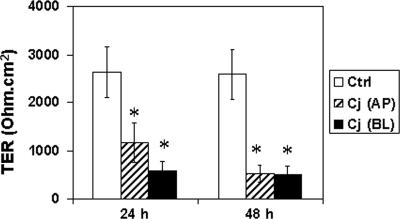
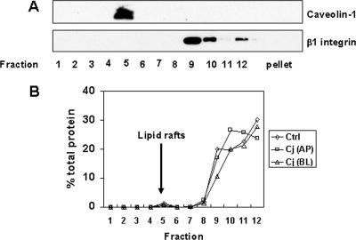
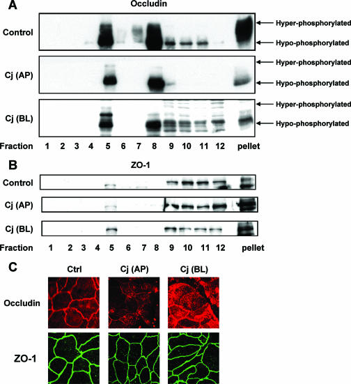
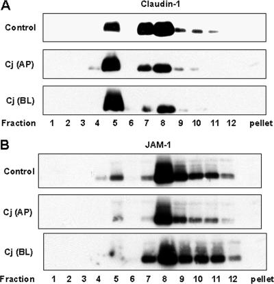
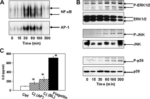
Campylobacter jejuni is a leading cause of human enterocolitis and is associated with postinfectious complications, including irritable bowel syndrome and Guillain-Barré syndrome. However, the pathogenesis of C. jejuni infection remains poorly understood. Paracellular pathways in intestinal epithelial cells are gated by intercellular junctions (tight junctions and adherens junctions), providing a functional barrier between luminal microbes and host immune cells in the lamina propria. Here we describe alterations in tight junctions in intestinal epithelial monolayers following C. jejuni infection. Apical infection of polarized T84 monolayers caused a time-dependent decrease in transepithelial electrical resistance (TER). Immunofluorescence microscopy revealed a redistribution of the tight junctional transmembrane protein occludin from an intercellular to an intracellular location. Subcellular fractionation using equilibrium sucrose density gradients demonstrated decreased hyperphosphorylated occludin in lipid rafts, Triton X-100-soluble fractions, and the Triton X-100-insoluble pellet following apical infection. Apical infection with C. jejuni also caused rapid activation of NF-kappaB and AP-1, phosphorylation of extracellular signal-regulated kinase, Jun N-terminal protein kinase, and p38 mitogen-activated protein kinases, and basolateral secretion of the CXC chemokine interleukin-8 (IL-8). Basolateral infection with C. jejuni caused a more rapid decrease in TER, comparable redistribution of tight-junction proteins, and secretion of more IL-8 than that seen with apical infection. These results suggest that compromised barrier function and increased chemokine expression contribute to the pathogenesis of C. jejuni-induced enterocolitis.


Figures

FIG. 1.
FIG. 1.
Reduced transepithelial electrical resistance in T84 cells infected with C. jejuni. Confluent T84 cells grown on permeable supports were infected with C. jejuni (Cj) at an MOI of 10 from either the apical (AP) or basolateral (BL) aspect. TER (ohm · cm2) was determined at 0, 24, and 48 h postinfection. Data (means ± standard deviations [error bars]; n = 12 for each group) were analyzed by paired Student t test, and an asterisk indicates significant differences between control (Ctrl) and infected cell samples at P < 0.05. In addition, there was significant difference (P < 0.05) between apical versus basolateral infection at 24 h.
FIG. 2.
FIG. 2.
Characterization of lipid rafts isolated by sucrose density gradient centrifugation. Lipid rafts were isolated by Triton X-100 detergent extraction and sucrose gradient centrifugation as described in Materials and Methods. Twelve 1-ml aliquots were collected from sucrose gradients, and proteins associated with the pellet were extracted with 1% SDS. (A) Equal amounts of protein from each fraction were analyzed by immunoblotting, probing with antibodies against caveolin-1 or β1 integrin, markers for lipid rafts and the basolateral membrane, respectively. Caveolin-1 was localized in fraction 5, which coincided with the light-scattering band visualized in the same fraction. The immunoblot shown is representative of the immunoblots for three experiments. (B) Protein concentration was determined by Dc protein assay and expressed as a percentage of total protein. The averages of three experiments collected from untreated control (Ctrl) cells and cells infected with C. jejuni (Cj) from either the apical (AP) or basolateral (BL) aspect are shown.
FIG. 3.
FIG. 3.
Effects of C. jejuni on the TJ proteins occludin and ZO-1. T84 cells were infected with C. jejuni (Cj) at an MOI of 10 from either the apical (AP) or basolateral (BL) aspect for 24 h. Cell lysates were subjected to sucrose density gradient centrifugation and analyzed by immunoblotting, probing with antibodies against occludin (A) or ZO-1 (B). Occludin migrated as hyper- and hypophosphorylated forms on SDS-polyacrylamide gels. The blots shown are representative of the blots for three separate studies. (C) Control cells and T84 cells infected with C. jejuni for 24 h were fixed by paraformaldehyde immediately after infection. Cells were incubated with antibodies against occludin or ZO-1 followed by species-specific secondary antibodies coupled to Alexa 555 (red, occludin) and Alexa 488 (green, ZO-1), respectively. The micrographs shown are representative of the micrographs for three experiments.
FIG. 4.
FIG. 4.
Equilibrium sucrose density gradient analysis of the TJ proteins claudin-1 and JAM-1. Cell lysates were prepared from control cells and T84 cells infected with C. jejuni (Cj) from either the apical (AP) or basolateral (BL) aspect for 24 h. Cell lysates were subjected to sucrose density gradient centrifugation. Equal amounts of protein from each fraction were analyzed by immunoblotting for either claudin-1 (A) or JAM-1 (B). The immunoblots presented are representative of the immunoblots for three experiments.
FIG. 5.
FIG. 5.
Proinflammatory responses induced by C. jejuni infection. T84 cells were infected with C. jejuni at an MOI of 10 for the times indicated. (A) The binding of NF-κB and AP-1 to oligonucleotides containing respective consensus sequences was assayed by electromobility shift assay. The results shown are representative of the results for three experiments. (B) The cytosolic fraction prepared from control and C. jejuni-infected cells was analyzed for Erk1/2, JNK, and p38 phosphorylation. Immunoblots were stripped and reprobed for total Erk1/2, JNK, and p38. The immunoblots shown are representative of the immunoblots for three experiments. P-ERK1/2, phosphorylated ERK1/2. (C) T84 cells were infected with C. jejuni (Cj) at an MOI of 10 for 24 h from either the apical (AP) or basolateral aspect (BL) (n = 9). In parallel cultures, cells were exposed to 100 ng/ml of S. enterica serovar Typhimurium flagellin from the basolateral aspect for 24 h (n = 6). Media in the lower chamber were assayed for IL-8 by enzyme-linked immunosorbent assays. The mean ± standard deviation (error bar) values for IL-8 secretion are shown. An asterisk indicates significant difference (P < 0.05) between control (Ctrl) and infected cell samples. In addition, there was a significant difference (P < 0.05) in IL-8 secretion between apical and basolateral C. jejuni infection.

References

    1. Abreu, M. T., M. Fukata, and M. Arditi. 2005. TLR signaling in the gut in health and disease. J. Immunol. 174:4453-4460. - PubMed
    1. Abreu, M. T., P. Vora, E. Faure, L. S. Thomas, E. T. Arnold, and M. Arditi. 2001. Decreased expression of Toll-like receptor-4 and MD-2 correlates with intestinal epithelial cell protection against dysregulated proinflammatory gene expression in response to bacterial lipopolysaccharide. J. Immunol. 167:1609-1616. - PubMed
    1. Akira, S., and K. Takeda. 2004. Toll-like receptor signalling. Nat. Rev. Immunol. 4:499-511. - PubMed
    1. Amieva, M. R., R. Vogelmann, A. Covacci, L. S. Tompkins, W. J. Nelson, and S. Falkow. 2003. Disruption of the epithelial apical-junctional complex by Helicobacter pylori CagA. Science 300:1430-1434. - PMC - PubMed
    1. Andersen-Nissen, E., K. D. Smith, K. L. Strobe, S. L. Barrett, B. T. Cookson, S. M. Logan, and A. Aderem. 2005. Evasion of Toll-like receptor 5 by flagellated bacteria. Proc. Natl. Acad. Sci. USA 102:9247-9252. [Epub ahead of print.] - PMC - PubMed

Publication types

MeSH terms

LinkOut - more resources