. 2006 Oct;26(20):7420-9.
doi: 10.1128/MCB.00597-06. Epub 2006 Aug 5.

AML1/RUNX1 phosphorylation by cyclin-dependent kinases regulates the degradation of AML1/RUNX1 by the anaphase-promoting complex

Affiliations

AML1/RUNX1 phosphorylation by cyclin-dependent kinases regulates the degradation of AML1/RUNX1 by the anaphase-promoting complex

Joseph R Biggs et al. Mol Cell Biol. 2006 Oct.

Abstract

AML1 (RUNX1) regulates hematopoiesis, angiogenesis, muscle function, and neurogenesis. Previous studies have shown that phosphorylation of AML1, particularly at serines 276 and 303, affects its transcriptional activation. Here, we report that phosphorylation of AML1 serines 276 and 303 can be blocked in vivo by inhibitors of the cyclin-dependent kinases (CDKs) Cdk1 and Cdk2. Furthermore, these residues can be phosphorylated in vitro by purified Cdk1/cyclin B and Cdk2/cyclin A. Mutant AML1 protein which cannot be phosphorylated at these sites (AML1-4A) is more stable than wild-type AML1. AML-4A is resistant to degradation mediated by Cdc20, one of the substrate-targeting subunits of the anaphase-promoting complex (APC). However, Cdh1, another targeting subunit used by the APC, can mediate the degradation of AML1-4A. A phospho-mimic protein, AML1-4D, can be targeted by Cdc20 or Cdh1. These observations suggest that both Cdc20 and Cdh1 can target AML1 for degradation by the APC but that AML1 phosphorylation may affect degradation mediated by Cdc20-APC to a greater degree.


Figures
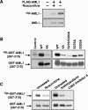
FIG. 1.
FIG. 1.
Phosphorylation of AML1 is inhibited by CDK inhibitors. (A) 293T cells were transfected with full-length FLAG-AML1 and then split into two samples. One sample was treated for approximately 20 h with 30 μM roscovitine. Nontransfected cells served as a control. During the last 4 to 6 h of roscovitine treatment, all samples were labeled with [32P]orthophosphate. The FLAG-AML1 was then immunoprecipitated with anti-FLAG agarose and used for Western blotting with anti-AML1 antibodies and for autoradiography. (B) Wild-type GST-AML1(267-315), GST-AML1(267-315)-4A (serines 276, 293, and 303 and threonine 300 mutated to alanine), GST-AML1(267-315)-S276/S303A (serines 276 and 303 mutated to alanine), or GST-AML1(267-315) with single mutations (serine 276 to alanine and serine 303 to alanine) were transfected into 293T cells, as indicated above the lanes. The far left lane shows nontransfected control cells. All cells were labeled with 32P, and the GST-AML1(267-315) was isolated using glutathione agarose. The GST-AML1(267-315) was then used for Western blotting with anti-GST antibodies and for autoradiography. (C) 293T cells were transfected with GST-AML1(267-315), split into separate samples, and then treated with CDK inhibitors as indicated above the lanes. Sixteen hours after treatment with CDK inhibitors, all samples were labeled using [32P]orthophosphate (CDK inhibitor concentrations were maintained during labeling). The GST-AML1(267-315) was pulled down using glutathione agarose and subjected to Western blotting with anti-GST antibodies and for autoradiography.
FIG. 2.
FIG. 2.
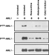
Phosphorylation of AML1 serines 276 and 303 is blocked by CDK inhibitors. 293T cells were transfected with FLAG-AML1 and then divided. One sample was left untreated, while the others were treated with 100 μM roscovitine, 50 μM alsterpaullone, or 50 μM Cdk2 inhibitor II, as indicated above the lanes. Nontransfected 293T cells were used as a control. Twenty hours after treatment, all samples were lysed and FLAG-AML1 was immunoprecipitated with anti-FLAG-agarose. Each sample of immunoprecipitated FLAG-AML1 was then used for Western blotting with anti-serine 276-phosphorylated AML1 (PS278-AML1) antibodies, anti-serine 303-phosphorylated AML1 (PS303-AML1) antibodies, and anti-AML1 antibodies.
FIG. 3.
FIG. 3.
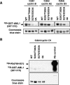
Cdk1/cyclin B, Cdk2/cyclin A, and Cdk6/cyclin D phosphorylate GST-AML1(267-315) in vitro, but Cdk4/cyclin D1 does not. (A) Wild-type GST-AML1B(267-315), GST-AML1B(267-315) with serine 293 and threonine 300 mutations, GST-AML1B(267-315) with serine 276 and 303 mutations, and GST-AML1B(267-315) with mutations of serines 276, 293, and 303 and threonine 300(4A) were expressed in 293T cells and bound to glutathione agarose. The agarose was incubated in kinase buffer with [γ-32P]ATP and either purified active Cdk1/cyclin B, Cdk2/cyclin A2, or Cdk6/cyclin D, as indicated above the lanes. After incubation for 10 min at 30°C, the GST-AML1(267-315) was boiled off the agarose in SDS sample buffer and run on an SDS polyacrylamide gel, which was stained with Coomassie blue to verify loading of proteins. The gel was then dried and used for autoradiography. (B) GST-AML1 substrates were incubated as above with purified active Cdk4/cyclin D1 along with a fusion protein containing amino acids 769 to 921 of the retinoblastoma protein as a positive control for Cdk4/cyclin D1 activity. All substrates were then analyzed as described for panel A.
FIG. 4.
FIG. 4.
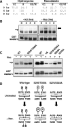
Cross talk between AML1 phosphorylation sites. (A) Cell cycle state of 293T cells synchronized with hydroxyurea or nocodazole. 293T cells were treated for 16 to 24 h with either 2 mM hydroxyurea or 0.1 mg/ml nocodazole or left untreated. Cells were collected immediately after removal of the hydroxyurea (HU) or nocodazole (Noc) (0 h) and after 4 and 8 h of culture. All cell samples were then fixed in ethanol, stained with propidium iodide, and analyzed by flow cytometry. The percentages of cells in the different phases of cell cycle are indicated. (B) Cells were treated as above and labeled with [32P]orthophosphate for 4 h before collection. After labeling, the GST-AML1(267-315) was isolated from cell lysates using glutathione agarose and used for Western blotting with anti-GST antibodies and for autoradiography. (C) 293T cells were transfected with wild-type or mutant GST-AML1(267-315) as indicated above the lanes. Some samples were treated for 16 to 20 h with 1 μg/μl nocodazole, as indicated above the lanes. During the last 4 to 6 h of nocodazole treatment, all samples were labeled with [32P]orthophosphate. After labeling, samples were analyzed as described for panel B. (D) A diagram of AML1 phosphorylation in 293T cells arrested in G2/M by nocodazole is shown. The upper left part of the diagram indicates that in unsynchronized cells, phosphorylation is detected only on serines 276 and 303 of GST-AML1(267-315). When the cells are arrested at G2/M by treatment with nocodazole, additional phosphorylation on serine 293 and/or threonine 300 is observed. The deletion of serine 293 and threonine 300 phosphorylation sites greatly enhances the phosphorylation at serines 276 and 303 of AML1. If serines 276 and 303 are mutated to alanine, the G2/M-specific phosphorylation at 293/300 does not occur (indicated at the right).
FIG. 5.
FIG. 5.
Mutation of AML1 phosphorylation sites to alanine increases cellular levels of AML1. (A) Lysates from control AEL-ΔR1 endothelial cells infected with empty MSCV-puro vector and cells stably expressing wild-type AML1, AML1B-4A mutant, or the phospho-mimic AML1B-4D mutant protein were used for Western blotting with anti-HA antibodies. Two independently infected pools expressing each type of AML1 were analyzed. Samples were stained with Ponceau solution after transfer to membranes to confirm approximately equal loading. (B) Total RNA was prepared from the pools of cells used to make the protein lysates analyzed above in panel A. The RNA was run on an agarose gel, and the 28S rRNA was stained with ethidium bromide to determine relative amounts of RNA in each lane. The RNA was then transferred to a membrane for Northern blotting with an AML1 probe. Protein lysates (C) and RNA (D) were prepared from NIH 3T3 cells infected with empty MSCV-puro vector and cells stably expressing wild-type AML1 and AML1B-4A mutant and analyzed as described for panels A and B. Equal loading of the NIH 3T3 protein lysate samples was confirmed by staining with Ponceau solution after transfer (not shown) and by Western blotting with antitubulin antibodies. Con, control.
FIG. 6.
FIG. 6.
Wild-type and phospho-mutant AML1 display different stabilities after cycloheximide treatment. (A) Endothelial cell lines expressing wild-type AML1, AML1-4A, or AML1-4D were treated for 0, 8, or 16 h with 20 μg/ml cycloheximide (CHX) as indicated above the lanes. Before lysis, the number of cells in each sample was determined, after which whole-cell lysates were prepared from each sample. Lysate from an equal number of cells was loaded into each lane, and the level of AML1 in each sample was determined by Western blotting with anti-AML1 antibodies. Ponceau staining of the membrane after transfer from the SDS gel was used to determine the amount of total protein in each sample. The relative amount of wild-type AML1, AML1-4A, or AML1-4D in each sample was calculated by densitometry, and the value was normalized to the amount of total protein (values are given beneath the upper panel). (B) Endothelial cells expressing HA-tagged wild-type AML1 were treated for 0, 8, or 20 h with 20 mg/ml cycloheximide (CHX) as indicated above the lanes. AML1 was then immunoprecipitated with anti-HA antibodies and analyzed by Western blotting with anti-serine 303-phosphorylated AML1 antibodies and anti-AML1 antibodies.
FIG. 7.
FIG. 7.
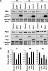
APC targeting subunit Cdc20 promotes the degradation of AML1 in a phosphorylation-dependent manner. (A) 293T cells stably expressing wild-type AML1, AML1B-4A mutant, or the phospho-mimic AML1B-4D mutant protein were transfected with either empty vector (control) or vector expressing Cdh1, Cdc20, or Skp2. At 24 to 48 h after transfection, cell lysates were prepared and used for Western blotting with anti-AML1 antibodies, followed by anti-HA antibodies to detect HA-tagged Cdh1 or Cdc20. Samples were stained with Ponceau solution after transfer to membranes to confirm approximately equal loading. (B) Three independent transfection experiments were performed as described for panel A using Cdh1, Cdc20, and Skp2. The quantity of wild-type AML1, AML1-4A, or AML1-4D was determined by densitometry of the bands visualized with the anti-AML1 antibodies. Differences in sample loading were corrected by measuring the intensity of the protein bands stained with Ponceau solution. The resulting values are presented graphically, with the amount of AML1 in the sample transfected with empty vector set at 1.0.
FIG. 8.
FIG. 8.
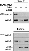
Cdc20 promotes the degradation of phosphorylated wild-type AML1 but not nonphosphorylated AML1. 293T cells were cotransfected with FLAG-tagged wild-type AML1 and either empty vector, Cdh1 expression vector, or Cdc20 expression vector, as indicated above the lanes. At 48 h after transfection, FLAG-AML1 was immunoprecipitated, and the immunoprecipitate was used for Western blotting with anti-serine 303-phosphorylated AML1 antibodies and anti-AML1 antibodies. Western blotting was also performed with samples of lysate, and anti-HA antibodies were used to detect Chd1 and Cdc20 expression.
FIG. 9.
FIG. 9.
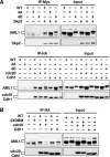
AML1 associates with Cdh1, Cdc20, and Skp2, and the AML1 destruction boxes are required for efficient association with Cdc20 but are not required for Cdh1. (A) 293T cells were transfected with the indicated expression plasmids and lysed in PBS-1 mM EDTA-0.5% Triton X-100. Excess AML1 expression vector was used for cotransfections to ensure overexpression of AML1 relative to Cdh1, Cdc20, or Skp2. This was done to make sure that induced degradation of AML1 was not observed and confused with any effects phosphorylation might have on the association of AML1 with Cdh1, Cdc20, or Skp2. A total of 200 μg of each sample was immunoprecipitated (IP) with the indicated antibodies, and immunoblotting was performed with anti-AML1 followed by anti-myc or anti-HA. Ten micrograms of each lysate was also resolved by SDS-PAGE and immunoblotting was performed as described above to determine the level of protein expression in every sample. (B) 293T cells were transfected with the indicated plasmids and the experiment performed as described above. DBM, destruction box mutant.
FIG. 10.
FIG. 10.
Diagram depicting cell cycle-dependent AML1 degradation by the APC/C or SCF complexes. Our data support the idea that AML1 phosphorylated at positions 276, 293, 300, and 303 is targeted for degradation by the APC-Cdc20 complex at early M phase. The APC-Cdh1 complex active during the late M and G1 phases is able to degrade AML1 independent of phosphorylation status. During reentry of the cells into S phase, the SCF-Skp2 complex slightly degrades phosphorylated AML1. This suggests a mechanism for the regulation of AML1 protein levels (and activity) during cell cycle progression.

Similar articles

Cited by

References

    1. Bain, J., H. McLauchlan, M. Elliott, and P. Cohen. 2003. The specificities of protein kinase inhibitors: an update. Biochem. J. 371:199-204. - PMC - PubMed
    1. Bernardin, F., and A. D. Friedman. 2002. AML1 stimulates G1 to S progression via its transactivation domain. Oncogene 21:3247-3252. - PubMed
    1. Bernardin-Fried, F., T. Kummalue, S. Leijen, M. I. Collector, K. Ravid, and A. D. Friedman. 2004. AML1/RUNX1 increases during G1 to S cell cycle progression independent of cytokine-dependent phosphorylation and induces cyclin D3 gene expression. J. Biol. Chem. 279:15678-15687. - PubMed
    1. Biggs, J. R., Y. Zhang, L. F. Peterson, M. Garcia, D. E. Zhang, and A. S. Kraft. 2005. Phosphorylation of AML1/RUNX1 regulates its degradation and nuclear matrix association. Mol. Cancer Res. 3:391-401. - PubMed
    1. Burns, C. E., D. Traver, E. Mayhall, J. L. Shepard, and L. I. Zon. 2005. Hematopoietic stem cell fate is established by the Notch-Runx pathway. Genes Dev. 19:2331-2342. - PMC - PubMed

Publication types

MeSH terms