. 2006 Oct;26(20):7760-71.
doi: 10.1128/MCB.00913-06.

Mutation in the Trapalpha/Ssr1 gene, encoding translocon-associated protein alpha, results in outflow tract morphogenetic defects

Affiliations

Mutation in the Trapalpha/Ssr1 gene, encoding translocon-associated protein alpha, results in outflow tract morphogenetic defects

K Mesbah et al. Mol Cell Biol. 2006 Oct.

Abstract

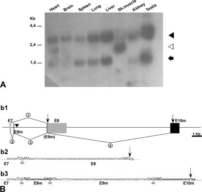
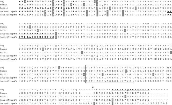
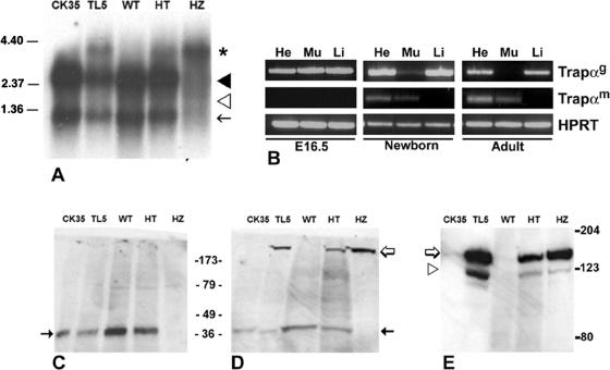
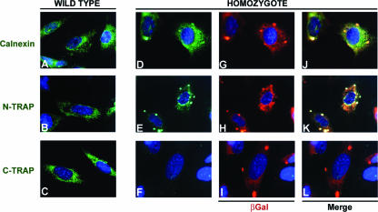
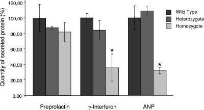
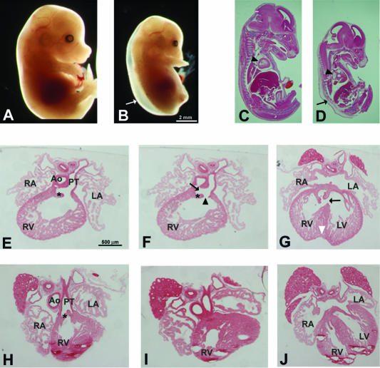
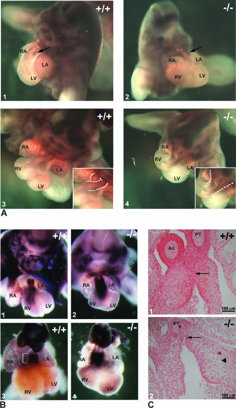
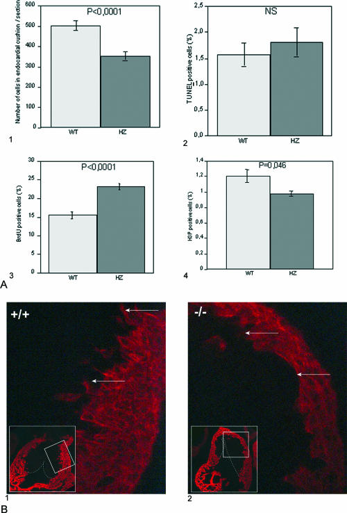
Translocon-associated protein complex (TRAP) is thought to be required for efficient protein-specific translocation across the endoplasmic reticulum membrane. We created a mutation in the Trapalpha gene that leads to the synthesis of a truncated TRAPalpha protein fused to ShBle-beta-galactosidase. Analysis of Trapalpha cDNAs reveals that among three different messenger RNAs expressed in the mouse, one of them encodes a slightly larger protein that differs in its C-terminal end. This mRNA, specific for skeletal muscle and heart, is only expressed after birth. Homozygous Trapalpha mutant pups die at birth, likely as a result of severe cardiac defects. Indeed, the septation of the proximal part of the outflow tract is absent, resulting in a double-outlet right ventricle. Studies of protein secretion in transfected embryonic fibroblasts reveal that the TRAP complex does not function properly in homozygous mutant cells and confirm, in vivo, the involvement of TRAP in substrate-specific translocation. Our results provide the first in vivo demonstration that a member of the TRAP complex plays a crucial role in mammalian heart development and suggest that TRAPalpha could be involved in translocation of factors necessary for maturation of endocardial cushions.


Figures

FIG. 1.
FIG. 1.
Characterization of Trapα messengers. (A) Northern blot analysis of mouse adult tissues (commercial membrane; Clontech) using a 638-bp probe encompassing the 5′ region of Trapα cDNA. Three different messengers of 2.7 kb (black arrowhead), 1.8 kb (white arrowhead), and 1.2 kb (black arrow) are shown. (B) 3′ end of the three different Trapα cDNAs. Sk.muscle, skeletal muscle. (b1) Schematic representation of 3′-genomic region encompassing the last exons of the Trapα gene. Angular lines indicate splicing events, E7 and E8 (gray) correspond to the last two exons of both the 2.7-kb and 1.2-kb messengers; E8m and E10m (black) are the two exons specific to the 1.8-kb mRNA. E9m is identical to the E8 coding region. A filled inverted triangle indicates the position of the vector insertion. (b2 and b3) Sequences of the 3′-end coding region of the different mRNAs. (b2) Sequence of 2.7-kb and 1.2-kb mRNAs resulting from splicing event 1 in b1. (b2) Sequence of 1.8 kb resulting from splicing events 2, 3, and 4 shown in b1. Black arrows indicate the ends of the coding regions. In b2 and b3 the three nucleotides at each extremity of the exons are in large uppercase letters and the stop codons are underlined. Numbers below the sequence indicate nucleotide positions in the cDNA sequences.
FIG. 2.
FIG. 2.
Sequence comparison between mouse TRAPα proteins and their human, dog, and rabbit orthologs. Shaded amino acids display some differences according to the species. The signal peptide region is in italics, while the transmembrane region is boxed. Underlined sequences correspond to the immunopeptides used to produce N- and C-terminal antibodies. The point of junction between TRAPα and ShBle-β-galactosidase in the hybrid protein synthesized in TL5 is indicated by a filled inverted triangle. Mouse TRAPαg corresponds to the protein expressed in all tissues but muscles; mouse TRAPαm corresponds to the muscle-specific protein. Protein sequence accession numbers from the SwissProt database: human, P43307; dog, P16967; rabbit, PS3815.
FIG. 3.
FIG. 3.
Trapα mRNA and protein expression in ES cells, embryos, or adult mouse cells. (A) Northern blot analysis of poly(A)+ RNAs isolated from ES cells (CK35, TL5) and from E15.5 embryos (WT, HT, HZ) hybridized with a 638-bp Trapα probe (see the legend to Fig. 1). Two messengers of 2.7 kb (black arrowhead) and 1.2 kb (black arrow) are expressed in WT and HT forms, both in ES cells and embryos; they are absent in the HZ embryos. A larger band of 4.2-kb mRNA (asterisk) is observed in HT or HZ embryos. It represents the fusion between TRAPα and ShBle-β-galactosidase mRNAs. The 1.8-kb RNA (white arrowhead) is expressed neither in ES cells nor in embryos. (B) RT-PCR of poly(A)+ RNA purified from heart (He), muscle (Mu), and liver (Li) from E16.5, newborn, or adult mouse cells. Trapαg corresponds to both 2.7- and 1.2-kb RNAs, while Trapαm is specific for the 1.8-kb RNAs. HPRT, hypoxanthine phosphoribosyltransferase. (C to E) Western blot analysis of ES cells and E15.5 embryos. Antibodies raised against C- or N-terminal TRAPα (C and D) or against β-galactosidase (E) were used. (C) C-terminal antibody revealed a protein of 34 kDa (black arrow) in wild-type (CK35, WT) and heterozygous (TL5, HT) samples but not in HZ samples. (D) N-terminal antibody binds the 34-kDa bands (black arrow) in WT and HT samples; in addition, it reveals a larger band of >123 kDa (white arrow) present in TL5, HT, and HZ samples but not in the WT. That larger band is the only one present in the HZ embryos. (E) Antibody directed against β-galactosidase binds to an identical band of >123 kDa (white arrow) in TL5, HT, and HZ samples, demonstrating that it corresponds to the TRAPα-ShBle-β-galactosidase fusion protein. The fainter band of 123 kDa, recognized by β-galactosidase antibody, corresponds to ShBle-β-galactosidase translated from the ATG present in the gene trap construct (white arrowhead).
FIG. 4.
FIG. 4.
Subcellular localization of TRAPα and TRAPα-ShBle-β-galactosidase fusion protein in WT and HZ embryonic fibroblasts. Immunofluorescence staining was performed using Calnexin antibody (green) (A, D, and J), TRAPα N-terminal antibody (N-TRAP; green) (B, E, and K), TRAPα C-terminal antibody (C-TRAP; green) (C, F, and L), and β-galactosidase antibody (βGal; red) (G to I). TRAPα N-terminal antibody stains both WT (B) and HZ (E) samples and reveals that TRAPα is localized in the same region as calnexin (A and D), thus, in the ER membrane. TRAPα C-terminal antibody stains WT cells (C) but does not recognize any protein in the HZ cells (F). β-Galactosidase colocalizes with calnexin (G and J) and TRAPα (H and K), revealing that the fusion protein is also inserted in the ER membrane. However, in HZ cells, the distribution of TRAPα-ShBle-β-galactosidase fusion protein in the membrane, revealed by either the N-terminal (E) or β-galactosidase antibody (G and H), is different from the distribution of TRAPα in wild-type cells (B); indeed, aggregates are clearly visible.
FIG. 5.
FIG. 5.
Secretion of proteins by embryonic fibroblasts after cotransfection of vectors expressing enhanced GFP and either preprolactin, IFN-γ, or ANP. After Western blot analysis, quantitation of secreted proteins was normalized to GFP content. The quantities of proteins secreted by either WT, HT, or HZ cells were plotted as, respectively, black, gray, and light gray bars (each bar corresponds to the mean for three independent samples). For each protein the secretion by wild-type fibroblasts was defined as 100%. Error bars represent the standard errors of the means; *, P < 0.05.
FIG. 6.
FIG. 6.
Trapα mutants are retarded and exhibit a strong cardiac phenotype. (A and B) The E14.5 HZ TL5 mutant (B) is growth retarded compared to a WT littermate (A). In addition, the HZ embryo displays an important subcutaneous edema (B, arrow). (C and D) Sagittal sections of E15.5 embryos reveal that in the TL5 mutant (D), the organs are proportionally smaller compared to those of the WT (C), except for the heart (arrowheads), which displays an important dilatation. (E to J) In serial transverse sections through E15.5 HZ (E to G) and WT (H to J) hearts, a partial separation of the aorta (Ao) and the pulmonary trunk (PT) is evident in the mutant embryo (F, arrow). This defect gives rise to a DORV (F, arrowhead) and an interventricular septal defect (G, arrow). RA, right atrium; LA, left atrium; RV, right ventricle; LV, left ventricle. Asterisks in panels E, F, and H show the semilunar valves. The white arrowhead in panel G points to the indentation in the ventricle apex.
FIG. 7.
FIG. 7.
Development of the outflow tract. (A) Neural crest cells visualized after whole-mount in situ hybridization on the trunk of E12.5 embryos using a plexin A2 riboprobe. Comparison between a TL5 mutant (A2 and 4) and a WT littermate (A1 and 3) reveals that the extent of migration of the crest cells is similar (A1 and 2, arrows). However, the route followed by the cells is very different in TL5 HZ embryos (compare the dashed lines in the insets in A3 and 4). (B) Endocardial cushion maturation as indicated by expression of periostin, which marks endocardial cells after EMT. Periostin expression in E12.5 WT (B1) and mutant (B2) or E13.5 WT (B3) and mutant (B4) embryos shows that endocardial cushions of the mutant had undergone EMT but that the staining is broader in the mutant at E13.5, suggesting an absence of maturation of the cushions (compare brackets). (C) Sections through OFT of E12.5 embryos revealed that the cushions have already fused in the WT (arrow in C1), while they are still separated in the mutant (arrow in C2). In addition, detachment of the cushions from the myocardial layer is frequently observed in the mutant (arrowhead in C2). Ao, aorta; PT, pulmonary trunk; RA, right atrium; LA, left atrium; RV, right ventricle; LV, left ventricle.
FIG. 8.
FIG. 8.
Endocardial cushion development. (A) Proliferation and apoptotic death of mesenchymal cells in endocardial cushions. (A1) Total number of mesenchymal cells in endocardial cushion of WT or HZ TL5 E12.5 OFT. Cells were counted on sagittal paraffin sections stained by hematoxylin (13 sections per genotype). Apoptotic death was analyzed using TUNEL (A2), while proliferation was evaluated by either BrdU (A3) or phosphohistone H3 (H3P; A4) immunostaining. In each case, results were expressed as the mean of the ratios of the number of positive cells to the total number of mesenchymal cells per section. NS, no significant difference between WT and HZ embryos was observed by TUNEL assay (six sections per genotype). An important increase in the number of BrdU-positive cells (five sections per genotype) and a small but significant (P < 0.05) decrease in the number of phosphohistone H3-positive cells (four sections per genotype) were revealed in HZ embryos. These results were confirmed with two different WT or HZ Trapα mutant embryos. Statistical significance of difference was assessed by Student's t testing. (B) Myocardialization of the endocardial cushions. Myosin heavy chain immunostaining with MF20 antibody on frontal sections of OFT in E12.5 embryos revealed that while migration of myocardial cells is clearly observed in the WT (B1, arrows), it has not yet started in the mutant (B2, arrows).

Similar articles

Cited by

References

    1. Azhar, M., J. Schultz Jel, I. Grupp, G. W. Dorn II, P. Meneton, D. G. Molin, A. C. Gittenberger-de Groot, and T. Doetschman. 2003. Transforming growth factor beta in cardiovascular development and function. Cytokine Growth Factor Rev. 14:391-407. - PMC - PubMed
    1. Bartram, U., D. G. M. Molin, L. J. Wisse, A. Mohamad, L. P. Sandford, T. Doestchman, C. P. Speer, R. E. Poelmann, and A. C. Gittenberger-de Groot. 2001. Double-outlet right ventricle and overriding tricuspid valve reflect disturbances of looping, myocardialization, endocardial cushion differentiation, and apoptosis in TGFbeta(2)-knockout mice. Circulation 103:2745-2752. - PubMed
    1. Beckmann, R., D. Bubeck, R. Grassucci, P. Penczek, A. Verschoor, G. Blobel, and J. Frank. 1997. Alignment of conduits for the nascent polypeptide chain in the ribosome-Sec61 complex. Science 278:2123-2126. - PubMed
    1. Brown, C. B., L. Feiner, M. M. Lu, J. Li, X. Ma, A. L. Webber, L. Jia, J. A. Raper, and J. A. Epstein. 2001. PlexinA2 and semaphorin signaling during cardiac neural crest development. Development 128:3071-3080. - PubMed
    1. Camus, A., C. Kress, C. Babinet, and J. Barra. 1996. Unexpected behavior of a gene trap vector comprising a fusion between the Sh ble and lacZ genes. Mol. Reprod. Dev. 45:255-263. - PubMed

Publication types

MeSH terms