. 2006 Oct 9;175(1):33-9.
doi: 10.1083/jcb.200603073. Epub 2006 Oct 2.

Kidney failure in mice lacking the tetraspanin CD151

Affiliations

Kidney failure in mice lacking the tetraspanin CD151

Norman Sachs et al. J Cell Biol. .

Abstract

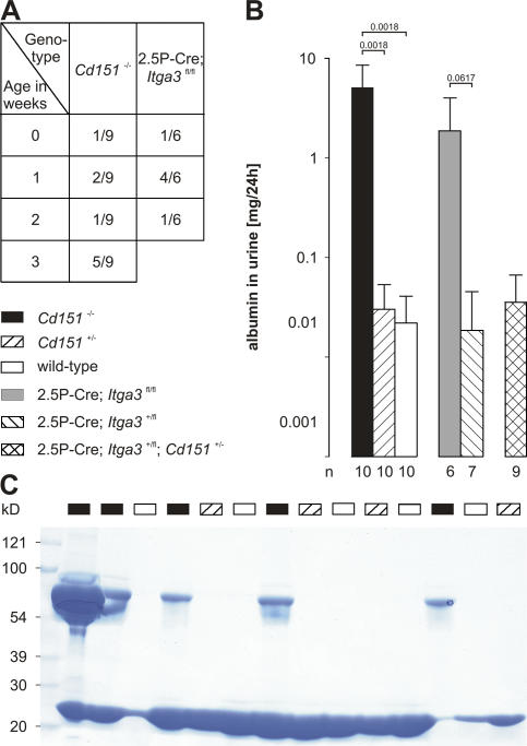
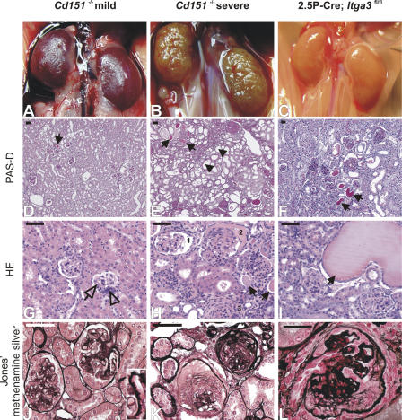
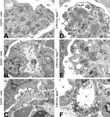
The tetraspanin CD151 is a cell-surface molecule known for its strong lateral interaction with the laminin-binding integrin alpha3beta1. Patients with a nonsense mutation in CD151 display end-stage kidney failure associated with regional skin blistering and sensorineural deafness, and mice lacking the integrin alpha3 subunit die neonatally because of severe abnormalities in the lung and kidney epithelia. We report the generation of Cd151-null mice that recapitulate the renal pathology of human patients, i.e., with age they develop massive proteinuria caused by focal glomerulosclerosis, disorganization of the glomerular basement membrane, and tubular cystic dilation. However, neither skin integrity nor hearing ability are impaired in the Cd151-null mice. Furthermore, we generated podocyte-specific conditional knockout mice for the integrin alpha3 subunit that show renal defects similar to those in the Cd151 knockout mice. Our results support the hypothesis that CD151 plays a key role in strengthening alpha3beta1-mediated adhesion in podocytes.


Figures

Figure 1.
Figure 1.
Massive albuminuria in Cd151−/− and 2.5P-Cre; Itga3fl/fl mice. (A) Age at which proteinuria was first detected by SDS-PAGE in Cd151 −/− and 2.5P-Cre; Itga3 fl/fl mice. (B) 24-h urinary albumin concentration in 6-wk-old mice as determined by competitive ELISA. Each bar represents the arithmetic mean ± the SD with P values, as determined by homoscedastic two-tailed t test, shown above it and the number of analyzed animals shown underneath. (C) Coomassie brilliant blue–stained gel showing the presence of albumin in the urine of Cd151 −/− mice in a group of three litters (1 μl urine per lane; age, 6 wk).
Figure 2.
Figure 2.
Glomerular and tubular abnormalities in mutant mice. Kidneys of Cd151 −/− and 2.5P-Cre; Itga3 fl/fl mice reveal mild (left column) and severe (middle and right columns) renal pathology. Although the mildly affected kidneys of a 3-mo-old Cd151 −/− mouse show a relatively smooth and intact surface (A), histological examination reveals protein casts (D, arrow), inconspicuous interstitial fibrosis, mesangial hypercellularity (G, open arrowhead), and mild glomerular sclerosis (G, open arrow). The GBM shows partial thickening and spike formation (J, inset). Severe lesions of another 3-mo-old Cd151 −/− mouse are characterized by a granular surface (B) caused by extensive glomerular collapsing sclerosis, periglomerular fibrosis, and focal interstitial inflammation and fibrosis (E, H, and K). The glomeruli show segmental (H, 1) and global (H, 2) attachment of glomerular tufts to Bowman's capsule, parietal epithelial crescents (H, 2), and hypercellularity, capillary collapse, and sclerosis (H, 3). Proximal tubuli show cystic dilation (E, arrowheads) and degeneration, as well as protein casts indicative of proteinuria (E and H, arrows). Silver staining indicates excessive GBM damage in some glomeruli (K). Severely affected kidneys of a 6-wk-old 2.5P-Cre; Itga3 fl/fl mouse reveal prominent protein casts and some extremely dilated proximal tubuli (F and I, arrows). The glomeruli are partially sclerosed and show substantial damage of the GBM (L). Bars, 50 μm.
Figure 3.
Figure 3.
Electron micrographs reveal GBM abnormalities and FP effacement in kidneys of mildly affected Cd151−/− and 2.5P-Cre; Itga3fl/fl, but not 2.5P-Cre; Itga3+/fl, mice. (A) Glomeruli of Cd151 −/− mice (3-mo-old) show abnormal capillary loops characterized by GBM thickening and loss of the regular pattern of podocyte FPs. (B) Peripheral glomerular capillary with a luminal thrombocyte and loss of fenestrations (arrowhead). The GBM is irregularly thickened and shows protrusions toward the capsular space (*). Podocyte FPs are partially lost (arrows). (C) Irregular GBM showing lamination, thickening, and protrusions (*). The endothelium is swollen and fenestrations are partially lost (arrowheads), as are podocyte FPs (arrows). Glomeruli of 6-wk-old 2.5P-Cre; Itga3 +/fl mice do not show abnormalities (D), whereas all glomeruli of 2.5P-Cre; Itga3 fl/fl mice exhibit severe structural changes (E and F). (E) Abnormally thickened GBM with protrusions throughout the glomerulus. (F) Podocyte FPs are completely lost (arrows) and GBM protrusions (*) are present along the entire length of the capillary loops. Solid lines point to normal structures (GBM and FPs), whereas dotted lines indicate abnormal thickening of the GBM and effacement of FPs. Bc, Bowman's capsule; Cs, capsular space; E, erythrocyte; Ec, endothelial cell; Fp, foot process; Lb, lamina basalis (GBM); Pdc, podocyte; T, thrombocyte; V, vascular lumen. Bars: (A, D, and E) 10 μm; (B, C, and F) 2 μm.
Figure 4.
Figure 4.
Histological analysis of Cd151−/− and 2.5P-Cre; Itga3fl/fl mice. Immunofluorescence analysis of kidneys from 3-mo-old wild-type (left column), mildly affected, and severely affected Cd151 −/− (second and third column), and 6-wk-old 2.5P-Cre; Itga3 +/fl and 2.5P-Cre; Itga3 fl/fl mice (columns four and five). The respective proteins are shown in green and red, yielding yellow upon colocalization. Nuclei are counterstained with TOPRO (blue). (A–C) Up-regulation of tenascin-C in the mildly affected, and of fibulin-2 in the severely affected, Cd151 −/− glomeruli. (D and E) 2.5P-Cre; Itga3 +/fl glomeruli show little presence of tenascin-C, whereas both tenascin-C and fibulin-2 are present in glomeruli of 2.5P-Cre; Itga3 fl/fl mice. (F–I) Integrin α3 and α6 are localized in glomeruli and tubules, respectively, in both wild-type and mildly affected Cd151 −/− kidneys, as well as in the kidneys of 2.5P-Cre; Itga3 +/fl mice. (J) Hardly any α3 is present in the glomeruli of 2.5P-Cre; Itga3 fl/fl mice, whereas α6 is normally distributed in the tubules. (K and N) Staining of nidogen shows regular GBMs in both wild-type and 2.5P-Cre; Itga3 +/fl mice. (L and M) Unusually strong nidogen staining of the GBM of peripheral capillary loops in both mildly and severely affected Cd151 −/− kidneys (arrowheads). Note the extracapillary accumulation of nidogen (arrow) and massive capillary collapse (β1 staining) in the severely affected kidneys. (O) Nidogen also accumulates in the capsular space of 2.5P-Cre; Itga3 fl/fl mice (arrow). Glomeruli are not collapsed, but show an abnormally thick GBM (arrowhead) with protrusions (open arrow). (P–T) Podocin is present along the glomerular tuft in wild-type, mildly affected Cd151 −/−, and 2.5P-Cre; Itga3 +/fl mice, but absent in severely affected Cd151 −/− and 2.5P-Cre; Itga3 fl/fl mice. Staining of laminin-10 shows sclerosed glomeruli in Cd151 −/− mice (R) and GBM thickening (arrowhead) with protrusions (open arrow) in 2.5P-Cre; Itga3 fl/fl mice (T). (U–W) Nephrin is lost in some glomeruli of mildly affected Cd151 −/− mice, but in all glomeruli of the severely affected Cd151 −/− mice. There is massive up-regulation of the collagen α2 (IV) in the severe Cd151 −/− phenotype (W) which is further characterized by hypercellularity in both the interstitium and glomeruli (C, H, M, R, and W; blue channel). (X and Y) Nephrin is also reduced in 2.5P-Cre; Itga3 fl/fl mice as compared with the conditional heterozygote, whereas the collagen α2 (IV) is hardly up-regulated. Insets K–O, green channel (nidogen); Inset T, red channel (laminin-10). Bar, 50 μm.

References

    1. Assad, L., M.M. Schwartz, I. Virtanen, and V.E. Gould. 1993. Immunolocalization of tenascin and cellular fibronectins in diverse glomerulopathies. Virchows Arch. B Cell Pathol. Incl. Mol. Pathol. 63:307–316. - PubMed
    1. Borradori, L., and A. Sonnenberg. 1999. Structure and function of hemidesmosomes: more than simple adhesion complexes. J. Invest. Dermatol. 112:411–418. - PubMed
    1. Boucheix, C., and E. Rubinstein. 2001. Tetraspanins. Cell. Mol. Life Sci. 58:1189–1205. - PMC - PubMed
    1. Cowin, A.J., D. Adams, S.M. Geary, M.D. Wright, J.C. Jones, and L.K. Ashman. 2006. Wound healing is defective in mice lacking tetraspanin CD151. J. Invest. Dermatol. 126:680–689. - PMC - PubMed
    1. DiPersio, C.M., K.M. Hodivala-Dilke, R. Jaenisch, J.A. Kreidberg, and R.O. Hynes. 1997. α3β1 integrin is required for normal development of the epidermal basement membrane. J. Cell Biol. 137:729–742. - PMC - PubMed

Publication types