. 2006 Oct 10;103(41):15056-61.
doi: 10.1073/pnas.0607286103. Epub 2006 Oct 2.

Structural basis for understanding oncogenic p53 mutations and designing rescue drugs

Affiliations

Structural basis for understanding oncogenic p53 mutations and designing rescue drugs

Andreas C Joerger et al. Proc Natl Acad Sci U S A. .

Abstract

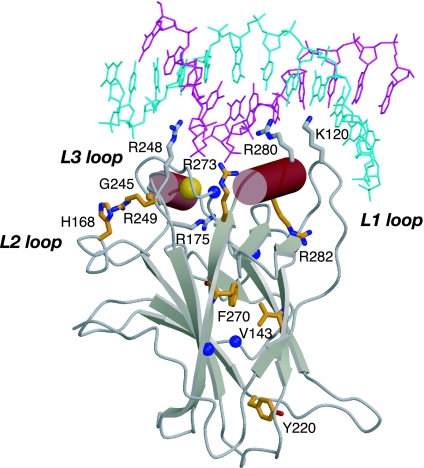
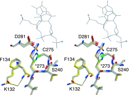
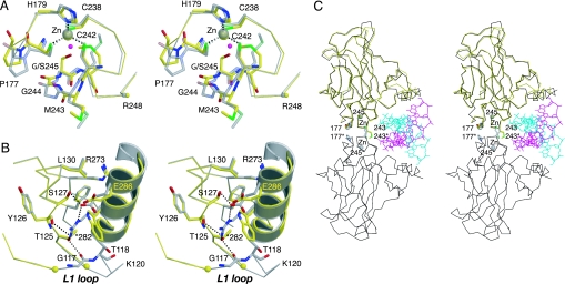
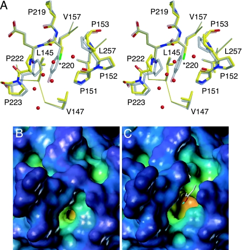
The DNA-binding domain of the tumor suppressor p53 is inactivated by mutation in approximately 50% of human cancers. We have solved high-resolution crystal structures of several oncogenic mutants to investigate the structural basis of inactivation and provide information for designing drugs that may rescue inactivated mutants. We found a variety of structural consequences upon mutation: (i) the removal of an essential contact with DNA, (ii) creation of large, water-accessible crevices or hydrophobic internal cavities with no other structural changes but with a large loss of thermodynamic stability, (iii) distortion of the DNA-binding surface, and (iv) alterations to surfaces not directly involved in DNA binding but involved in domain-domain interactions on binding as a tetramer. These findings explain differences in functional properties and associated phenotypes (e.g., temperature sensitivity). Some mutants have the potential of being rescued by a generic stabilizing drug. In addition, a mutation-induced crevice is a potential target site for a mutant-selective stabilizing drug.


Conflict of interest statement

The authors declare no conflict of interest.

Figures

Fig. 1.
Fig. 1.
Structure of the p53 core domain bound to consensus DNA (5). The two strands of bound consensus DNA are shown in blue and magenta. The bound zinc ion is displayed as a golden sphere. Cancer mutation sites that were structurally studied in this work and earlier work (16) are shown in orange. The blue spheres indicate the location of the mutation sites in the superstable quadruple mutant M133L/V203A/N239Y/N268D (T-p53C).
Fig. 2.
Fig. 2.
Crystal structure of T-p53C-R273C. Stereoview of the mutation site in the structure of T-p53C-R273C [Protein Data Bank (PDB) ID code 2J20, yellow] superimposed on the structure of T-p53C (PDB ID code 1UOL, orange), T-p53C-R273H (PDB ID code 2BIM, gray), and DNA-bound wild type (PDB ID code 1TSR, light gray). One strand of bound DNA in the vicinity of Arg-273 is shown as a gray line, with small spheres indicating the continuation of the DNA backbone.
Fig. 3.
Fig. 3.
Structural effects of the cancer mutations G245S and R282W. (A) Stereoview of the zinc-binding region of T-p53C-G245S (PDB ID code 2J1Y, molecule A, yellow) superimposed on the structure of T-p53C (PDB ID code 1UOL, molecule A, gray). The Cα atom of the DNA-contact residue Arg-248 is depicted as a small sphere in the color of the corresponding chain. A structural water molecule that is present in T-p53C but displaced by the side chain of Ser-245 in T-p53C-G245S is shown as a magenta sphere. (B) Stereoview of the loop–sheet–helix motif in the crystal structure of T-p53C-R282W (PDB ID code 2J21, molecule A, yellow) superimposed on T-p53C (PDB ID code 1UOL, molecule A, gray). Stabilizing interactions mediated via Arg-282 in T-p53C are shown as dotted lines. Residues 117–121 of T-p53C-R282W were disordered. The Cα atoms on both sides of the chain break are highlighted by small yellow spheres. (C) Cα trace of a wild-type core domain dimer bound to a DNA half-site (PDB ID code 2AC0, black) (10). The two strands of bound DNA are shown in blue and magenta. T-p53C-G245S, shown in yellow, is superimposed on one of the two monomers. Selected residues at the core–core domain interface are highlighted.
Fig. 4.
Fig. 4.
Crystal structure of T-p53C-Y220C. (A) Stereoview of the mutation site at the periphery of the β-sandwich in T-p53C-Y220C (PDB ID code 2J1X, molecule A, yellow) superimposed on the structure of T-p53C (PDB ID code 1UOL, molecule A, gray). Several water molecules close to Cys-220 in T-p53C-Y220C that fill the cleft created by the mutation are shown as red spheres. (B) Molecular surface of T-p53C around Tyr-220. (C) Molecular surface of T-p53C-Y220C. The view is the same as in B. The position of the side chain of Tyr-220 in T-p53C is shown as a stick model.
Fig. 5.
Fig. 5.
Crystal structures of T-p53C-V143A and T-p53C-F270L. (A) Stereoview of the structure of T-p53C-V143A (PDB ID code 2J1W, yellow) superimposed on T-p53C (PDB ID code 1UOL, gray). All residues in the hydrophobic core of the β-sandwich within a 4.5-Å radius of the Val-143 side chain in T-p53C are shown. (B) Stereoview of the structure of T-p53C-F270L (PDB ID code 2J1Z, yellow) superimposed on T-p53C (PDB ID code 1UOL, gray). All residues within a 6-Å radius of the Phe-270 side chain in T-p53C are shown.

References

    1. Vogelstein B., Lane D, Levine AJ. Nature. 2000;408:307–310. - PubMed
    1. Vousden KH, Lu X. Nat Rev Cancer. 2002;2:594–604. - PubMed
    1. Olivier M, Eeles R, Hollstein M, Khan MA, Harris CC, Hainaut P. Hum Mutat. 2002;19:607–614. - PubMed
    1. Beroud C, Soussi T. Hum Mutat. 2003;21:176–181. - PubMed
    1. Cho Y, Gorina S, Jeffrey PD, Pavletich NP. Science. 1994;265:346–355. - PubMed

Publication types

MeSH terms

Substances