. 2006 Oct 10;103(41):15085-90.
doi: 10.1073/pnas.0603057103. Epub 2006 Oct 2.

Androgen receptor as a licensing factor for DNA replication in androgen-sensitive prostate cancer cells

Affiliations

Androgen receptor as a licensing factor for DNA replication in androgen-sensitive prostate cancer cells

Ivan V Litvinov et al. Proc Natl Acad Sci U S A. .

Abstract

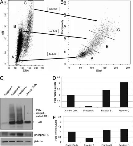
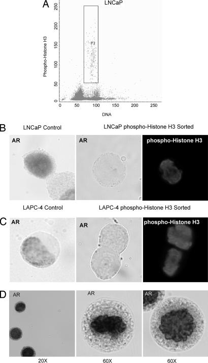
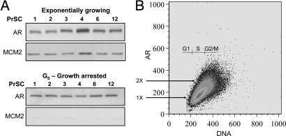
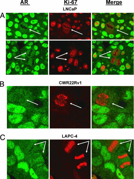
Androgen receptor (AR) protein expression and function are critical for survival and proliferation of androgen-sensitive (AS) prostate cancer cells. Besides its ability to function as a transcription factor, experimental observations suggest that AR becomes a licensing factor for DNA replication in AS prostate cancer cells and thus must be degraded during each cell cycle in these cells to allow reinitiation of DNA replication in the next cell cycle. This possibility was tested by using the AS human prostate cancer cell lines, LNCaP, CWR22Rv1, and LAPC-4. These studies demonstrated that AR levels fluctuate both within and between various phases of the cell cycle in each of these AS lines. Consistent with its licensing ability, AR is degraded during mitosis via a proteasome-dependent pathway in these AS prostate cancer cells. In contrast, proteasome-dependent degradation of AR during mitosis is not observed in AR-expressing but androgen-insensitive human prostate stromal cells, in which AR does not function as a licensing factor for DNA replication. To evaluate mitotic degradation of AR in vivo, the same series of human AS prostate cancers growing as xenografts in nude mice and malignant tissues obtained directly from prostate cancer patients were evaluated by dual Ki-67 and AR immunohistochemistry for AR expression in mitosis. These results document that AR is also down-regulated during mitosis in vivo. Thus, AS prostate cancer cells do not express AR protein during mitosis, either in vitro or in vivo, consistent with AR functioning as a licensing factor for DNA replication in AS prostate cancer cells.


Conflict of interest statement

The authors declare no conflict of interest.

Figures

Fig. 1.
Fig. 1.
AR protein expression during cell cycling of AS prostate cancer cells. (A) AR protein is expressed strongly in CWR22Rv1, LNCaP, and LAPC-4, is very weakly expressed in human PrSCs, and is not expressed in PC3 and DU145 cells. (BD) Dual-parameter FACS analyses for AR expression and DNA fluorescence analysis in PC3 (B), LNCaP (C), and CWR22Rv1 (D) prostate cancers cells. Results are presented as linear scale dot plots with DNA fluorescence (i.e., cell cycle stage) on the x axis and AR staining intensity on y axis. All analyzed cells were grown in culture in the presence of 1.0 nM R1881. PC3 expression pattern was used to set up a gate to identify specific (i.e., detectable) vs. nonspecific (i.e., nondetectable) staining for AR. This gate was then applied to all subsequent FACS analyses.
Fig. 2.
Fig. 2.
AR protein decreases in mitosis in AS prostate cancer cells. (A) Exponentially growing LNCaP cells in the presence of 1 nM R1881 were analyzed by dual-parameter FACS analysis, and subsequent fractions of cells were sorted on the basis of AR expression and DNA fluorescence. (B) Forward-side scatter analysis of LNCaP cells shows that fraction B cells are significantly larger and more complex than fraction A cells, and fraction C cells are significantly larger and more complex than fraction B cells. (CE) Western blot analyses and quantitation of AR and phospho-Rb protein expression in sorted A, B, and C fractions and exponentially growing LNCaP control cells. Quantitation of Western blot analysis is presented in D [folds of AR levels (normalized per actin)] and E [folds of phospho-Rb levels (normalized per actin)].
Fig. 3.
Fig. 3.
AR protein is not expressed in mitotic AS prostate cancer cells. (A) Flow sorting of exponentially growing LNCaP cells positive for phospho-histone H3 expression to select cells in mitosis. (B and C) Immunocytochemical analysis of AR expression in exponentially growing (control) LNCaP and LAPC-4 prostate cancer cells compared with phospho-histone H3+ (i.e., mitotic) sorted cells. Compared with exponentially growing (i.e., nonmitotic) AR+ LNCaP and LAPC-4 control cells (B Left and C Left), AR expression was undetectable in phospho-histone H3+ prostate cancer cells (B Center and Right and C Center and Right). (D) LNCaP cells were treated with MG-132 proteasome inhibitor for 6 h in the presence of R1881, flow sorted for phospho-histone H3 expression, and analyzed for AR expression by immunocytochemistry. Proteasome inhibition resulted in strong AR expression during mitosis.
Fig. 4.
Fig. 4.
AR expression during cell cycling of androgen-insensitive prostate stromal cells. (A) Normal human PrSCs (PrSC cells nos. 1–4, 6, and 12) were exponentially growing or arrested in G0 in the presence of androgen by contact inhibition. Protein lysates were immunoblotted for the expression of AR and the DNA replication licensing factor MCM2. (B) Dual-parameter FACS analysis of proliferating PrSC cell no. 6 shows that AR levels fluctuate from 1× to 2× throughout the cell cycle.
Fig. 5.
Fig. 5.
AR protein levels are down-regulated during mitosis in AS prostate cancer LNCaP (A), CWR22Rv1 (B), and LAPC-4 (C) xenografts grown in intact male nude mice. In these studies, tumor tissue sections were stained for AR (green) and the proliferation marker Ki-67 (red). Ki-67 is a well established marker for proliferation that is expressed in all parts of cell cycle except in proliferatively quiescent G0 or early G1 phases. Importantly, Ki-67 reacts with an interchromatin network during mitosis, thereby “painting” the chromosomes (36) and making it easy to identify mitotic cells (arrows). Consistent with the in vitro data, AR expression was greatly decreased in cells undergoing mitosis in LNCaP, CWR22Rv1, and LAPC-4 prostate cancer xenografts.

References

    1. Jemal A, Murray T, Ward E, Samuels A, Tiwari RC, Ghafoor A, Feuer EJ, Thun MJ. CA Cancer J Clin. 2005;55:10–30. - PubMed
    1. Shah RB, Mehra R, Chinnaiyan AM, Shen R, Ghosh D, Zhou M, Macvicar GR, Varambally S, Harwood J, Bismar TA, et al. Cancer Res. 2004;64:9209–9216. - PubMed
    1. van Bokhoven A, Varella-Garcia M, Korch C, Johannes WU, Smith EE, Miller HL, Nordeen SK, Miller GJ, Lucia MS. Prostate. 2003;57:205–225. - PubMed
    1. Litvinov IV, De Marzo AM, Isaacs JT. J Clin Endocrinol Metab. 2003;88:2972–2982. - PubMed
    1. Haag P, Bektic J, Bartsch G, Klocker H, Eder IE. J Steroid Biochem Mol Biol. 2005;96:251–258. - PubMed

Publication types

Substances