

Visual-evoked potentials to onset of chromatic red-green and blue-yellow gratings in Parkinson's disease never treated with L-dopa
- PMID: 17016154
- PMCID: PMC3703931
- DOI: 10.1097/01.wnp.0000216127.53517.4d
Visual-evoked potentials to onset of chromatic red-green and blue-yellow gratings in Parkinson's disease never treated with L-dopa
Abstract
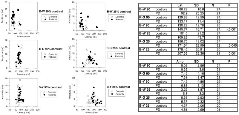
The differential dysfunction of chromatic and achromatic visual pathways in early Parkinson's disease (PD) was evaluated by means of visual-evoked potentials (VEPs) recorded in 12 patients (mean age 60.1 +/- 8.3 years; range 46 to 74 years) in the early stages of PD and not yet undergoing treatment with L-dopa, and in 12 age-matched controls. Visual stimuli were full-field (14 deg) equiluminant red-green (R-G), blue-yellow (B-Y), and black-white (B-W) sinusoidal gratings of two cycles per degree, presented in onset (300 milliseconds)--offset (700 milliseconds) mode, at two contrast (K) levels (90% and 25%). The VEP mean latencies were significantly more delayed in PD patients than in controls for chromatic than for luminance stimuli, in particular for B-Y stimuli of low contrast (K90%: B-W = 6.6 milliseconds, R-G = 3.34 milliseconds, B-Y = 15.48 milliseconds; K25%: B-W = 7.8 milliseconds, R-G = 14.8 milliseconds, B-Y = 28.9). Latencies of chromatic VEPs were more variable that achromatic VEP latencies in both normal subjects and PD patients. Therefore, the frequency of latency abnormalities (within 30%) was not significantly different for the three visual stimuli. Our results show that, in addition to achromatic VEPs, chromatic VEPs are impaired in early PD patients not yet undergoing L-dopa therapy, indicating an acquired color deficiency in these patients. The greater delay for the B-Y VEPs suggests a higher vulnerability of visual blue-cone pathway in the early stages of the disease. However, the overall sensitivity of chromatic VEPs in detecting early visual impairment in PD is comparable with that of achromatic VEPs.
Figures

Similar articles
-
Changes in pattern electroretinograms to equiluminant red-green and blue-yellow gratings in patients with early Parkinson's disease.J Clin Neurophysiol. 2003 Sep-Oct;20(5):375-81. doi: 10.1097/00004691-200309000-00010. J Clin Neurophysiol. 2003. PMID: 14701999
-
Equiluminant red-green and blue-yellow VEPs in multiple sclerosis.J Clin Neurophysiol. 2001 Nov;18(6):583-91. doi: 10.1097/00004691-200111000-00010. J Clin Neurophysiol. 2001. PMID: 11779973
-
Chromatic pattern-reversal electroretinograms (ChPERGs) are spared in multiple system atrophy compared with Parkinson's disease.Neurol Sci. 2006 Feb;26(6):395-401. doi: 10.1007/s10072-006-0522-1. Neurol Sci. 2006. PMID: 16601931 Free PMC article.
-
'Gamma' band oscillatory response to chromatic stimuli in volunteers and patients with idiopathic Parkinson's disease.Vision Res. 2009 Mar;49(7):726-34. doi: 10.1016/j.visres.2009.01.018. Epub 2009 Feb 14. Vision Res. 2009. PMID: 19232367 Free PMC article.
-
Chromatic visual evoked potentials: A review of physiology, methods and clinical applications.Prog Retin Eye Res. 2024 Jul;101:101272. doi: 10.1016/j.preteyeres.2024.101272. Epub 2024 May 16. Prog Retin Eye Res. 2024. PMID: 38761874 Review.
Cited by
-
Retina as a Model to Study In Vivo Transmission of α-Synuclein in the A53T Mouse Model of Parkinson's Disease.Methods Mol Biol. 2021;2224:75-85. doi: 10.1007/978-1-0716-1008-4_5. Methods Mol Biol. 2021. PMID: 33606207
-
Continuous Detection of Stimulus Brightness Differences Using Visual Evoked Potentials in Healthy Volunteers with Closed Eyes.Bioengineering (Basel). 2024 Jun 13;11(6):605. doi: 10.3390/bioengineering11060605. Bioengineering (Basel). 2024. PMID: 38927841 Free PMC article.
-
Color perception deficits in co-existing attention-deficit/hyperactivity disorder and chronic tic disorders.J Neural Transm (Vienna). 2008;115(2):235-9. doi: 10.1007/s00702-007-0817-2. Epub 2007 Sep 27. J Neural Transm (Vienna). 2008. PMID: 17896072
-
Visual Evoked Potentials for the Detection of Diabetic Retinal Neuropathy.Int J Mol Sci. 2023 Apr 17;24(8):7361. doi: 10.3390/ijms24087361. Int J Mol Sci. 2023. PMID: 37108524 Free PMC article. Review.
-
Retinal electrophysiology in central nervous system disorders. A review of human and mouse studies.Front Neurosci. 2023 Aug 2;17:1215097. doi: 10.3389/fnins.2023.1215097. eCollection 2023. Front Neurosci. 2023. PMID: 37600004 Free PMC article. Review.
References
-
- Adams AJ. Chromatic and luminosity processing in retinal disease. Am J Optom Physiol Opt. 1982;59:954–960. - PubMed
-
- Barbato L, Rinalduzzi S, Laurenti M, et al. Color VEPs in Parkinson’s disease. Electroencephalogr Clin Neurophysiol. 1994;92:169–172. - PubMed
-
- Birch J, Kolle RU, Kunkel M, et al. Acquired colour deficiency in patients with Parkinson’s disease. Vision Res. 1998;38:3421–3426. - PubMed
-
- Bodis-Wollner I, Marx MS, Mitra S, et al. Visual dysfunction in Parkinson’s disease. Loss in spatiotemporal contrast sensitivity. Brain. 1987;110:1675–1698. - PubMed
-
- Buttner T, Kuhn W, Muller T, et al. Chromatic and achromatic visual evoked potentials in Parkinson’s disease. Electroencephalogr Clin Neurophysiol. 1996;100:443–447. - PubMed
Publication types
MeSH terms
Grants and funding
LinkOut - more resources
Full Text Sources
Other Literature Sources
Medical
