

Review
doi: 10.1172/JCI29812.
Deiodinases: implications of the local control of thyroid hormone action
Affiliations
- PMID: 17016550
- PMCID: PMC1578599
- DOI: 10.1172/JCI29812
Item in Clipboard
Review
Deiodinases: implications of the local control of thyroid hormone action
J Clin Invest.
2006 Oct.
Abstract
The deiodinases activate or inactivate thyroid hormone, and their importance in thyroid hormone homeostasis has become increasingly clear with the availability of deiodinase-deficient animals. At the same time, heightened interest in the field has been generated following the discovery that the type 2 deiodinase can be an important component in both the Hedgehog signaling pathway and the G protein-coupled bile acid receptor 1-mediated (GPBAR1-mediated) signaling cascade. The discovery of these new roles for the deiodinases indicates that tissue-specific deiodination plays a much broader role than once thought, extending into the realms of developmental biology and metabolism.
Figures
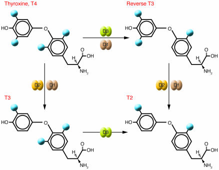
The reactions catalyzed by the deiodinases remove iodine moieties (blue spheres) from the phenolic (outer rings) or tyrosil (inner rings) rings of the iodothyronines. These pathways can activate T4 by transforming it into T3 (via D1 or D2) or prevent it from being activated by converting it to the metabolically inactive form, reverse T3 (via D1 or D3). T2 is an inactive product common to both pathways that is rapidly metabolized by further deiodination.
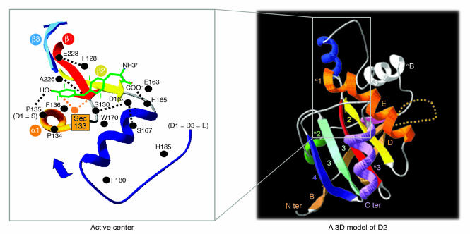
While the deiodinases have not yet been crystallized, protein modeling indicates that they share a common general structure composed of a single aminoterminal-anchoring segment, a short hinge region, and a thioredoxin fold–containing globular domain (9). A 3D model of the D2 globular domain is shown on the right. Letters and numbers shown indicate different β sheets and α-helices as previously reported (9). The orange dotted loop indicates the D2-specific segment that mediates interaction with the E3-ubiquitin ligase WSB-1 (28). The inset illustrates the active center, which contains the rare amino acid selenocysteine (Sec), which is critical for nucleophilic attack during the deiodination reaction. The residues that putatively interact with the T4 molecule (green) are also shown. Position 135, which in D2 and D3 is occupied by proline, is critical for enzyme kinetics. D2 and D3 have high affinity for their substrates and are not sensitive to inhibition by PTU. Replacement with serine, which is naturally found in D1, turns both D2 and D3 into low affinity and PTU-sensitive enzymes (9). C ter, C terminus; N ter, N terminus. Figure modified with permission from the Journal of Biological Chemistry (9) and Nature Cell Biology (28).
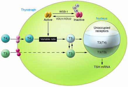
In the thyrotroph, TSH is subject to negative feedback by T3, which arrives in the nucleus from 2 distinct sources: plasma T3, illustrated as T3(T3); and plasma T4, which is then converted to T3 intracellularly via the D2 pathway, represented as T3(T4). The schematic includes the plasma membrane, which contains thyroid hormone transporters (indicated by the pink and red circles); the cytoplasm, containing the enzymes involved in thyroid hormone metabolism; and the nucleus, containing the TRs. D2 is represented in active form (yellow) and inactive form (red). Transition between active and inactive D2 is via ubiquitination and deubiquitination, reactions that are catalyzed by WSB-1 and VDU1/VDU2, respectively. As a result of ubiquitination, D2-mediated T4-to-T3 conversion occurs at variable rates, decreasing as serum T4 concentration increases (46). Ultimately, these processes determine nuclear TR saturation, which includes contributions from both T3(T3) and T3(T4) as indicated, with only a minor fraction of the TRs being unoccupied under normal conditions. Ub, Ubiquitin. Figure modified with permission from Endocrine Reviews (14).
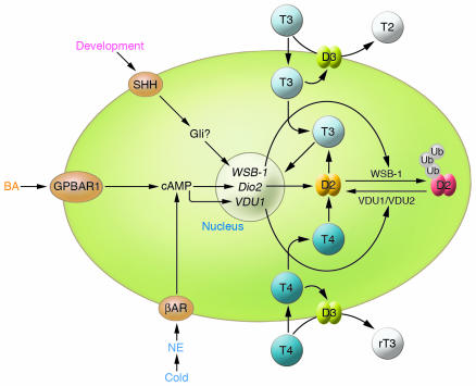
In D2-expressing cells, such as brown adipocytes, stimulation of D2 expression increases local T3 production, resulting in increased saturation of T3 receptors. This increase can be mediated by norepinephrine (NE) stimulation of β-adrenergic receptors (βARs), such as occurs during cold stimulation, or by bile acid–mediated (BA–mediated) stimulation of GPBAR1 (also known a TGR5). Both of these pathways activate cAMP production and stimulate Dio2 transcription. In brown fat, cAMP also promotes VDU1 expression, amplifying D2 induction via deubiquitination. Other signaling pathways can decrease D2 activity, resulting in relative local hypothyroidism. For example, the Hedgehog cascade decreases D2 activity by promoting WSB-1 expression and thus D2 ubiquitination, presumably via the Gli cascade. rT3, reverse T3; SHH, sonic hedgehog.
References
-
- Wu Y., Koenig R.J. Gene regulation by thyroid hormone. Trends Endocrinol. Metab. 2000;11:207–211. - PubMed
-
- Wondisford F.E. Thyroid hormone action: insight from transgenic mouse models. J. Investig. Med. 2003;51:215–220. - PubMed
-
- Yen P.M., et al. Thyroid hormone action at the cellular, genomic and target gene levels. Mol. Cell. Endocrinol. 2006;246:121–127. - PubMed
-
- St. Germain D.L., Galton V.A. The deiodinase family of selenoproteins. Thyroid. 1997;7:655–668. - PubMed
-
- Bianco A.C., Salvatore D., Gereben B., Berry M.J., Larsen P.R. Biochemistry, cellular and molecular biology and physiological roles of the iodothyronine selenodeiodinases. Endocr. Rev. 2002;23:38–89. - PubMed
Publication types
MeSH terms
Substances
Grants and funding
LinkOut - more resources
Full Text Sources
Other Literature Sources
Medical
