. 2006 Oct;116(10):2682-94.
doi: 10.1172/JCI29218.

Coordinated epithelial NHE3 inhibition and barrier dysfunction are required for TNF-mediated diarrhea in vivo

Affiliations

Coordinated epithelial NHE3 inhibition and barrier dysfunction are required for TNF-mediated diarrhea in vivo

Daniel R Clayburgh et al. J Clin Invest. 2006 Oct.

Abstract

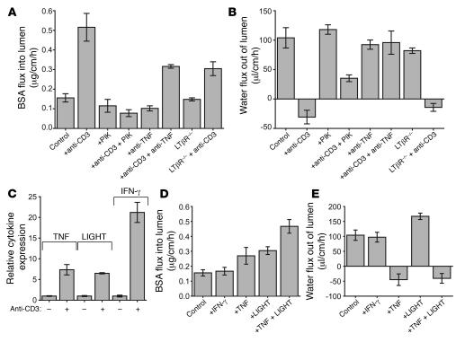
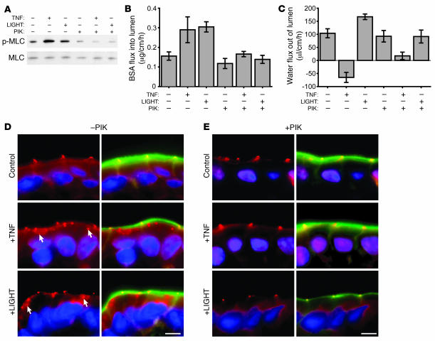
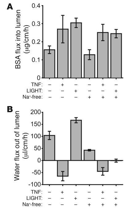
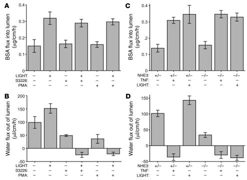
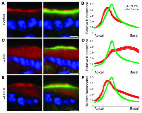
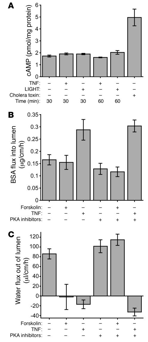
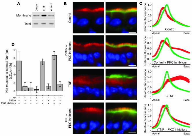
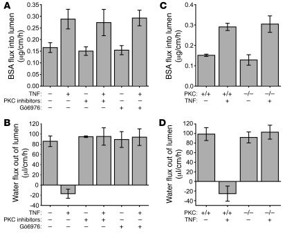
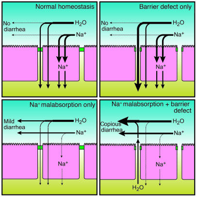
Acute T cell-mediated diarrhea is associated with increased mucosal expression of proinflammatory cytokines, including the TNF superfamily members TNF and LIGHT. While we have previously shown that epithelial barrier dysfunction induced by myosin light chain kinase (MLCK) is required for the development of diarrhea, MLCK inhibition does not completely restore water absorption. In contrast, although TNF-neutralizing antibodies completely restore water absorption after systemic T cell activation, barrier function is only partially corrected. This suggests that, while barrier dysfunction is critical, other processes must be involved in T cell-mediated diarrhea. To define these processes in vivo, we asked whether individual cytokines might regulate different events in T cell-mediated diarrhea. Both TNF and LIGHT caused MLCK-dependent barrier dysfunction. However, while TNF caused diarrhea, LIGHT enhanced intestinal water absorption. Moreover, TNF, but not LIGHT, inhibited Na+ absorption due to TNF-induced internalization of the brush border Na+/H+ exchanger NHE3. LIGHT did not cause NHE3 internalization. PKCalpha activation by TNF was responsible for NHE3 internalization, and pharmacological or genetic PKCalpha inhibition prevented NHE3 internalization, Na+ malabsorption, and diarrhea despite continued barrier dysfunction. These data demonstrate the necessity of coordinated Na+ malabsorption and barrier dysfunction in TNF-induced diarrhea and provide insight into mechanisms of intestinal water transport.


Figures

Figure 1
Figure 1. Administration of TNF and LIGHT induces barrier dysfunction similar to that caused by anti-CD3 injection, but only TNF causes net water secretion.
(A) In vivo perfusion assays show that anti-CD3 causes a large increase in BSA flux (P < 0.0001 versus control). The MLCK inhibitor PIK completely prevented this increased BSA flux, which was also attenuated by anti-TNF or use of LTβR–/– mice. (B) Anti-CD3 injection reverses water movement, from net water absorption in control animals to net water secretion (P < 0.0001). PIK restored water flow to net absorption, although absorption was still significantly less than in control animals. Anti-TNF completely restored water absorption after anti-CD3 injection. No significant water absorption or secretion was observed in LTβR–/– animals treated with anti-CD3 (P < 0.001). (C) Two hours following anti-CD3 injection, TNF, LIGHT, and IFN-γ mRNA were assessed in intestinal mucosa using quantitative real-time PCR. Anti-CD3 caused significant increases in transcripts for all 3 cytokines (P < 0.001). (D) Either TNF or LIGHT, but not IFN-γ, causes significant increases in BSA flux, though not as large as that caused by anti-CD3. Simultaneous injection of TNF and LIGHT led to a larger increase in BSA flux that resembled the increase following anti-CD3 injection. (E) While IFN-γ does not alter water movement, TNF reverses water movement from net water absorption to net water secretion in a manner similar to anti-CD3. In contrast, LIGHT caused an increase in water absorption (P = 0.03). Simultaneous TNF and LIGHT treatment caused water secretion similar to that caused by treatment with TNF alone.
Figure 2
Figure 2. TNF and LIGHT mediate epithelial barrier dysfunction via MLCK activity.
(A) Either TNF or LIGHT treatment increases MLC phosphorylation in jejunal epithelia. PIK prevented TNF- and LIGHT-mediated increases in MLC phosphorylation. (B) Either TNF or LIGHT causes increased BSA flux. PIK prevented increases in BSA flux after TNF or LIGHT treatment, indicating that epithelial barrier dysfunction after TNF or LIGHT treatment requires MLCK. (C) TNF causes net water secretion, while LIGHT increases net water absorption. PIK restored net absorption after TNF treatment, although a significant quantitative defect remained (P = 0.01). PIK reduced normal water absorption to control levels in animals treated with LIGHT. MLCK-mediated barrier dysfunction is therefore responsible for the changes in water flux due to LIGHT but does not completely account for changes due to TNF. (D) Immunofluorescent localization of occludin (red), F-actin (green), and nuclei (blue) in the jejunal epithelium shows that in control mice (top panels), occludin was localized to apical cell-cell junctions. After either TNF or LIGHT injection (middle and bottom panels), intracellular occludin inclusions were also present (white arrows). Scale bar: 5 μm. (E) Immunofluorescent localization of occludin (red), F-actin (green), and nuclei (blue) in the jejunal epithelium of mice treated with the MLCK inhibitor PIK demonstrates that occludin was localized exclusively at apical cell-cell junctions, indicating that PIK treatment prevented TNF- and LIGHT-mediated occludin internalization. Scale bar: 5 μm.
Figure 3
Figure 3. Induction of Na+ malabsorption reverses water flux in LIGHT-treated animals.
(A) Mice were injected with TNF, LIGHT, or vehicle and then perfused with solution containing Na+ or the Na+ substitute N-methyl- d-glucamine where indicated. Both TNF and LIGHT treatment caused a significant increase in BSA flux compared with control, and perfusion with Na+-free perfusate had no effect on the barrier dysfunction elicited by TNF or LIGHT. (B) When perfused with solution lacking Na+, control animals demonstrated a reduction in net water absorption (P = 0.03). TNF injection caused net water secretion regardless of the presence of Na+ in the perfusate. After LIGHT injection, perfusion with solution containing Na+ resulted in an increase in water absorption compared with that in control animals (P = 0.02), while perfusion with solution lacking Na+ led to complete ablation of water absorption (P = 0.008).
Figure 4
Figure 4. NHE3 inhibition coupled with LIGHT injection leads to net water secretion.
(A) LIGHT increases BSA flux into the perfused jejunal segment during in vivo perfusion assays regardless of treatment with S3226 or PMA. (B) LIGHT increases water absorption compared with that seen in control animals. Ten micromolar S3226 reduces water absorption in control animals (P = 0.02) and allows net water secretion in animals injected with LIGHT (P = 0.003). PMA also caused water malabsorption in control animals (P = 0.03) and net water secretion after LIGHT injection (P = 0.003). (C) Assay of NHE3+/– (+/–) and NHE3–/– (–/–) mice shows that both TNF and LIGHT cause increased BSA flux regardless of the presence of NHE3. (D) While NHE3+/– mice display normal water absorption, NHE3–/– mice have a significant quantitative defect (P = 0.02). TNF treatment of NHE3+/– or NHE3–/– mice caused net water loss similar to that in wild-type animals. LIGHT caused a nonsignificant increase in water absorption in NHE3+/– mice but induced net water secretion in NHE3–/– mice (P = 0.0003).
Figure 5
Figure 5. NHE3 is internalized after TNF treatment.
Immunofluorescent detection of NHE3 (red), F-actin (green), and nuclei (blue) in the jejunal epithelium 1 hour after injection of vehicle (A), 5 μg TNF (C), or 5 μg LIGHT (E) demonstrates that NHE3 is predominantly found in the epithelial brush border in both control and LIGHT-treated mice. After TNF treatment, much of this brush border staining was absent. Quantification of pixel intensities of multiple immunofluorescent images from control (B), TNF- (D), and LIGHT-treated (F) mice was performed. F-actin fluorescence (green) peaked at the perijunctional actomyosin ring in each set of images. NHE3 intensity (red) peaked just apical to the perijunctional actomyosin ring in controls and after LIGHT treatment, but this peak was abolished after TNF treatment, indicating that TNF leads to a significant loss of brush border NHE3. Scale bars: 5 μm.
Figure 6
Figure 6. cAMP signaling is not involved in TNF-mediated water secretion.
(A) Measurement of cAMP levels in the jejunal epithelium of mice 30 or 60 minutes after injection with either TNF or LIGHT shows that neither treatment increases epithelial cAMP. Cholera toxin is shown as a positive control. (B) BSA flux was measured in control animals, after the addition of 20 μM forskolin to the perfusion solution or after injection of 5 μg TNF. The PKA inhibitors KT5720 (500 nM) and myristoylated PKI (1 μM) were added to the perfusion solution as indicated. TNF, but not forskolin, significantly increased BSA flux. PKA inhibitors had no effect on BSA flux. (C) Both forskolin and TNF treatment resulted in net water secretion. PKA inhibitors restored net water absorption to control levels after forskolin treatment but had no effect on TNF-induced net water secretion, indicating that cAMP signaling is not involved in TNF-mediated water secretion.
Figure 7
Figure 7. PKCα activation occurs after TNF treatment and is responsible for Na+ malabsorption and NHE3 internalization.
(A) Membrane fractions were isolated from jejunal epithelia 1 hour after injection of vehicle, TNF, or LIGHT and immunoblotted for PKCα. TNF, but not LIGHT, increased PKCα expression in the membrane fraction, indicating that only TNF activates PKCα. (B) Immunofluorescent detection of NHE3 (red), F-actin (green), and nuclei (blue) in jejunal epithelia of control or TNF-treated mice perfused with the PKC inhibitors chelerythrine (10 μM) and bisindolylmaelimide (400 nM) demonstrates that the inhibitors do not change NHE3 localization in control animals. TNF caused internalization of brush border NHE3, but the PKC inhibitors prevented NHE3 loss after TNF treatment. (C) Line scan quantification of multiple immunofluorescent images confirms that PKC inhibitors alone do not alter NHE3 localization but that the inhibitors do preserve brush border NHE after TNF treatment. Thus, PKC is responsible for TNF-mediated NHE3 internalization. Scale bars: 5 μm. (D) Net mucosal-to-serosal Na+ flux was measured in Üssing chambers. Control animals demonstrated robust S3226-sensitive Na+ absorption. TNF reduced the S3226-sensitive component of net Na+ flux (P = 0.003). PKC inhibitors prevented TNF-mediated loss of S3226-sensitive Na+ absorption, indicating that PKC activity is necessary for TNF-mediated NHE3 downregulation.
Figure 8
Figure 8. PKC inhibition prevents TNF-mediated net water secretion.
(A) TNF increases BSA flux into the intestine during intestinal perfusion relative to that in control animals, and this is not affected by the general PKC inhibitors chelerythrine and bisindolylmaelimide or the PKCα inhibitor Gö6976 (10 μM). (B) TNF causes net water secretion. Either chelerythrine and bisindolylmaelimide or Gö6976 blocked TNF-mediated water secretion and restored net water absorption. (C) BSA flux is increased by TNF in PKCα-knockout mice (P = 0.005). (D) While TNF causes net water secretion in wild-type animals, net water absorption is maintained in PKCα–/– mice after TNF treatment.
Figure 9
Figure 9. A model of TNF-induced diarrhea.
Under normal conditions, most of the Na+ that enters the intestine is absorbed by the intestinal epithelium, producing an osmotic gradient that drives water absorption. When a small paracellular barrier defect is introduced, such as that occurring after LIGHT injection, water is still absorbed, as the osmotic gradient produced by Na+ transport remains intact; in fact, the increase in paracellular permeability may actually increase the amount of water absorbed. Alternatively, when Na+ transport alone is impaired, as occurs after NHE3 inhibition, water absorption is reduced, leading to mild malabsorptive diarrhea. When epithelial barrier dysfunction and Na+ malabsorption occur simultaneously, not only is water retained in the lumen, but additional water may egress through the paracellular spaces, contributing to a larger volume of diarrhea than occurs when a barrier defect is not present.

Comment in

Similar articles

Cited by

References

    1. Field M. Intestinal ion transport and the pathophysiology of diarrhea. J. Clin. Invest. 2003;111:931–943. doi: 10.1172/JCI200318326. - DOI - PMC - PubMed
    1. Hawker P.C., McKay J.S., Turnberg L.A. Electrolyte transport across colonic mucosa from patients with inflammatory bowel disease. Gastroenterology. 1980;79:508–511. - PubMed
    1. Clayburgh D.R., et al. Epithelial myosin light chain kinase-dependent barrier dysfunction mediates T cell activation-induced diarrhea in vivo. . J. Clin. Invest. 2005;115:2702–2715. doi: 10.1172/JCI24970. - DOI - PMC - PubMed
    1. Ferran C., et al. Cytokine-related syndrome following injection of anti-CD3 monoclonal antibody: further evidence for transient in vivo T cell activation. Eur. J. Immunol. 1990;20:509–515. - PubMed
    1. Musch M.W., et al. T cell activation causes diarrhea by increasing intestinal permeability and inhibiting epithelial Na+/K+-ATPase. J. Clin. Invest. 2002;110:1739–1747. doi: 10.1172/JCI200215695. - DOI - PMC - PubMed

Publication types

MeSH terms