. 2006 Oct;116(10):2808-16.
doi: 10.1172/JCI28310.

Hematopoietic stem cells proliferate until after birth and show a reversible phase-specific engraftment defect

Affiliations

Hematopoietic stem cells proliferate until after birth and show a reversible phase-specific engraftment defect

Michelle B Bowie et al. J Clin Invest. 2006 Oct.

Abstract

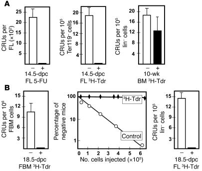
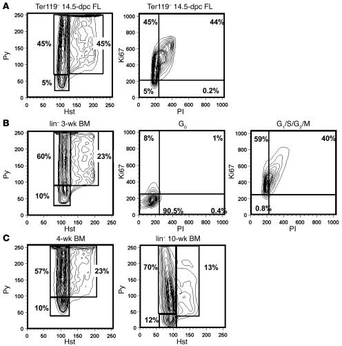
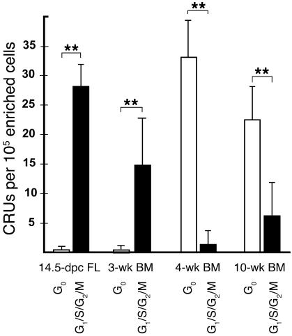
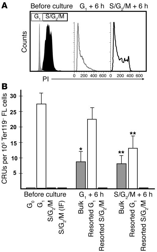
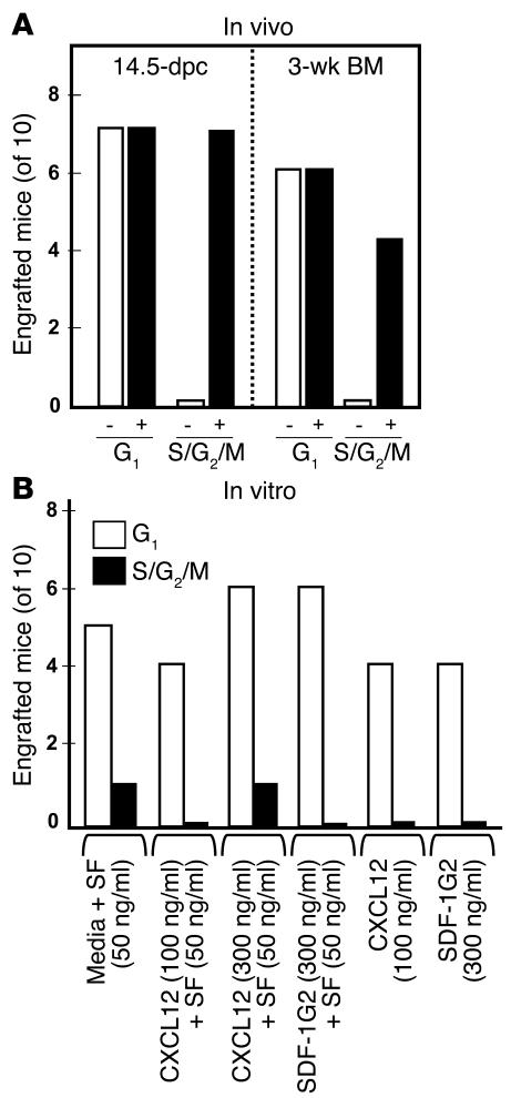
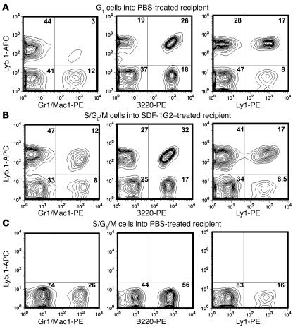
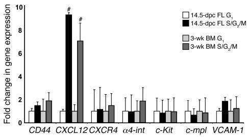
The regulation of HSC proliferation and engraftment of the BM is an important but poorly understood process, particularly during ontogeny. Here we show that in mice, all HSCs are cycling until 3 weeks after birth. Then, within 1 week, most became quiescent. Prior to 4 weeks of age, the proliferating HSCs with long-term multilineage repopulating activity displayed an engraftment defect when transiting S/G2/M. During these cell cycle phases, their expression of CXC chemokine ligand 12 (CXCL12; also referred to as stromal cell-derived factor 1 [SDF-1]) transiently increased. The defective engrafting activity of HSCs in S/G2/M was reversed when cells were allowed to progress into G1 prior to injection or when the hosts (but not the cells) were pretreated with a CXCL12 antagonist. Interestingly, the enhancing effect of CXCL12 antagonist pretreatment was exclusive to transplants of long-term multilineage repopulating HSCs in S/G2/M. These results demonstrate what we believe to be a new HSC regulatory checkpoint during development. They also suggest an ability of HSCs to express CXCL12 in a fashion that changes with cell cycle progression and is associated with a defective engraftment that can be overcome by in vivo administration of a CXCL12 antagonist.


Figures

Figure 1
Figure 1. All fetal HSCs are sensitive to cell cycle–specific drugs.
Cells from different mouse embryonic tissues were analyzed for CRU content either 16 hours after injection of the pregnant mother with 100 mg/kg 5-FU (+) or PBS (–) or after in vitro incubation of the cells for 16 hours with (+) or without (–) high–specific activity 3H-Tdr. (A) Shown are the effects of 5-FU injection on 14.5-dpc FL CRUs (left panel; data pooled from 3 independent experiments) as well as the effects of 3H-Tdr on 14.5-dpc FL CRUs (middle panel) and the lack of effect of 3H-Tdr on CRUs from adult (10-week-old) mice assessed in parallel (right panel; data pooled from 6 independent experiments). (B) The effects of 3H-Tdr on 18.5-dpc fetal BM (FBM) and FL CRUs (left and right panels; data pooled from 4 independent experiments). The middle panel shows the complete data set from the limiting dilution analysis of the 18.5-dpc fetal BM cells. **P < 0.001.
Figure 2
Figure 2. FACS profiles of the distribution of G0, G1, and S/G2/M cells in different lin populations.
(A) Representative FACS contour plot for 14.5-dpc Ter119 FL cells after staining with Hst and Py and for Ki67. (B) Representative FACS contour plot for lin 3-wk BM cells after staining with Hst and Py; sorted G0 cells after staining for Ki67 (>90% of the G0 cells showed no Ki67 expression); and sorted G1/S/G2/M cells after staining for Ki67 (>99% of the G1/S/G2/M cells expressed Ki67). (C) Representative FACS contour plots for lin 4- and 10-wk BM cells after staining with Hst and Py. Percentages indicate the proportion of total cells found within the indicated gene.
Figure 3
Figure 3. The cycling activity of CRUs is downregulated between 3 and 4 weeks of age.
Shown are the number of CRUs per 105 initial total viable cells. FL cells were depleted of Ter119+ cells; for the 3- and 4-wk BM cells, all lin+ cells except Mac1+ cells were removed; and for the 10-wk BM cells, all lin+ cells including Mac1+ cells were removed. Values are mean ± SEM from data pooled from at least 3 experiments per tissue. **P < 0.001.
Figure 4
Figure 4. Hst/Py-sorted HSCs display an absolute but transient S/G2/M engraftment defect.
(A) Ter119 14.5-dpc FL cells in G1/S/G2/M were fractionated into their component G1 and S/G2/M subsets, leaving a slight separation between them. Aliquots of the sorted subsets were then stained with PI as well as with Hst/Py (data not shown). The sorted cells were cultured for 6 hours and then stained again with PI. We found that during this 6-hour culture period, approximately one-third of the cells originally in G1 had progressed into S/G2/M, and a similar proportion of the cells originally in S/G2/M had progressed into G1. (B) CRUs per 105 initial Ter119 FL cells for G1 and S/G2/M fractions before and after 6 hours in culture. There was a 3.5-fold loss of CRUs when G1 cells were cultured for 6 hours, but no loss when the cultured cells were re-sorted for G1 cells (P = 0.36). Conversely, we detected a greater than 65-fold increase in the number of CRUs detected when CRUs in S/G2/M were cultured and a greater than 128-fold increase when the cultured cells were sorted for G1 cells. IF, intrafemoral. Values are mean ± SEM of results from at least 3 experiments. *P < 0.01, **P < 0.001 versus respective cell types before culture.
Figure 5
Figure 5. The engraftment defect of HSCs in S/G2/M is corrected by treatment of the host, but not the cells, with SDF-1G2.
(A) Effect of injecting prospective recipients 2 hours after irradiation and 2 hours prior to transplant with 10 ng/ml SDF-1G2 (+) or PBS (–). Starting equivalents of 4,000 G1 cells per recipient mouse or 12,000 S/G2/M cells per recipient mouse were similarly tested. Both 14.5-dpc FL and 3-wk BM HSCs in S/G2/M engrafted only when transplanted into SDF-1G2–treated recipients, whereas treated recipients were no more likely to be engrafted long-term by HSCs in G1 than were untreated recipients. Results are combined from 3 independent experiments. (B) Effect of in vitro treatment of sorted Ter119 FL cells in G1 or S/G2/M for 30 minutes at 37°C in serum-free medium plus the indicated additives on CRU detection. In vitro treatment had no significant effect on the number of mice that subsequently showed multilineage repopulation from starting cells in either G1 or S/G2/M. Results are combined from 3 independent experiments.
Figure 6
Figure 6. Donor-derived repopulation of SDF-1G2–treated mice.
Shown are representative FACS profiles of donor-specific cells detected after dual staining for the donor-type Ly5 allotype and various lineage-specific markers. (A) Example of a positively engrafted PBS-treated recipient of FL cells in G1. (B) Example of a positively engrafted SDF-1G2–treated recipient of FL cells in S/G2/M. (C) Example of a PBS-treated recipient of FL cells in S/G2/M that did not show donor-derived hematopoiesis. Numbers within graphs indicate the proportion of total cells found within the indicated gene.
Figure 7
Figure 7. Gene expression analysis of the G1 and S/G2/M subsets of highly purified linSca1+CD43+Mac1+ HSCs from 14.
-dpc FL and 3-wk BM. Gene expression in G1 was set as 1, and the fold change in transcript levels in the corresponding S/G2/M fraction is shown. Results are mean ± SEM of data from 2–3 biological replicates measured in triplicate. #P < 0.05 versus respective G1 samples.

Comment in

References

    1. Szilvassy S.J., Humphries R.K., Lansdorp P.M., Eaves A.C., Eaves C.J. Quantitative assay for totipotent reconstituting hematopoietic stem cells by a competitive repopulation strategy. Proc. Natl. Acad. Sci. U. S. A. 1990;87:8736–8740. - PMC - PubMed
    1. Harrison D.E., Astle C.M., Lerner C.1984. . Ultimate erythropoietic repopulating abilities of fetal, young adult, and old adult cells compared using repeated irradiation. J. Exp. Med. 160759–771. - PMC - PubMed
    1. Fleming W.H., et al. Functional heterogeneity is associated with the cell cycle status of murine hematopoietic stem cells. J. Cell Biol. 1993;122:897–902. - PMC - PubMed
    1. Leemhuis T., et al. Isolation of primitive human bone marrow hematopoietic progenitor cells using Hoechst 33342 and Rhodamine 123. Exp. Hematol. 1996;24:1215–1224. - PubMed
    1. Osawa M., Hanada K., Hamada H., Nakauchi H. Long-term lymphohematopoietic reconstitution by a single CD34-low/negative hematopoietic stem cell. Science. 1996;273:242–245. - PubMed

Publication types

MeSH terms