

Benign pulmonary metastasizing leiomvomatosis: case report and a review of the literature
- PMID: 17017666
- PMCID: PMC3890720
- DOI: 10.3904/kjim.2006.21.3.173
Benign pulmonary metastasizing leiomvomatosis: case report and a review of the literature
Abstract
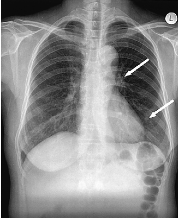
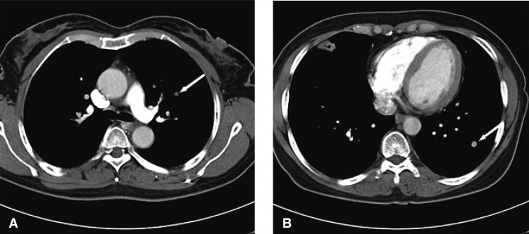
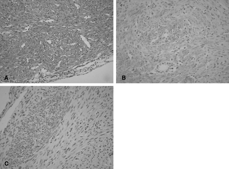
The authors report here on a case of a nearly asymptomatic 51-year-old Korean woman who was found to have diffuse, multiple nodules of the lungs on a routine chest radiograph. She had undergone hysterectomy 16 years previously for uterine myoma. An open lung biopsy revealed tumor that was composed of interlacing bundles of spindle cells with cigar shaped nucleus and eosinophilic myofibrils in the cytoplasm; consistent with multiple leiomyomas. The stains for SMA, desmin, MSA and Ki-67 were positive and the stain for c-kit was negative. The other stains for estrogen and progesterone receptor were positive. During the open lung biopsy procedure, all the nodules were excised. We report here on an interesting case of benign metastasizing leiomyoma (BML) in 51-year-old patient. To the best of our knowledge, this case showed the longest period of clinical progression in Korea. This is also one of a few cases in which curative excision was successfully performed.
Figures
References
-
- Arai T, Yasuda Y, Takay T, Shibayama M. Natural decrease of benign metastasizing leiomyoma. Chest. 2000;117:921–922. - PubMed
-
- Uchida T, Tokumaru T, Kojima H, Nakagawaji K, Imaizumi M, Abe T. A case of multiple leiomyomatous lesions of the lung: an analysis of flow cytometry and hormone receptors. Surg Today. 1992;22:265–268. - PubMed
-
- Hwang JK, Park KY, Park JW, Park JK, Jeong SH, Suh JB, Lee HK, Lee JW, Oh YH, Nam GH. A case of benign metastasizing leiomyoma in the lung. Tuberc Respir Dis. 2000;49:231–236.
-
- Kim YS, Kim EJ, Park CH, Park JS, Jee YK, Lee KY. A case of benign metastasizing pulmonary leiomyomatosis. Tuberc Respir Dis. 2002;53:190–195.
-
- Kang SA, Choi SI, Kim YA, Kim CJ, Yang DG, Kang JH, Kie JH, Hong YK, Lee SM. A case of benign metastasizing pulmonary leiomyoma. Tuberc Respir Dis. 2005;58:614–618.
Publication types
MeSH terms
LinkOut - more resources
Full Text Sources
Medical
Research Materials