

doi: 10.1186/1477-7819-4-70.
Thoracoscopic enucleation of a large esophageal leiomyoma using a three thoracic ports technique
Affiliations
- PMID: 17018158
- PMCID: PMC1599730
- DOI: 10.1186/1477-7819-4-70
Item in Clipboard
Thoracoscopic enucleation of a large esophageal leiomyoma using a three thoracic ports technique
World J Surg Oncol.
.
Abstract
Background: Video assisted thoracoscopic resection of an esophageal leiomyoma offers distinct advantages over an open approach. Many papers have described various techniques of thoracoscopic resection.
Case presentation: We describe a 32-year old man who presented with intermittent dysphagia. Imaging studies showed a large esophageal leiomyoma. He underwent thoracoscopic enucleation using a three thoracic-ports technique.
Conclusion: Thoracoscopic enucleation can be technically performed using a three thoracic-ports technique.
Figures
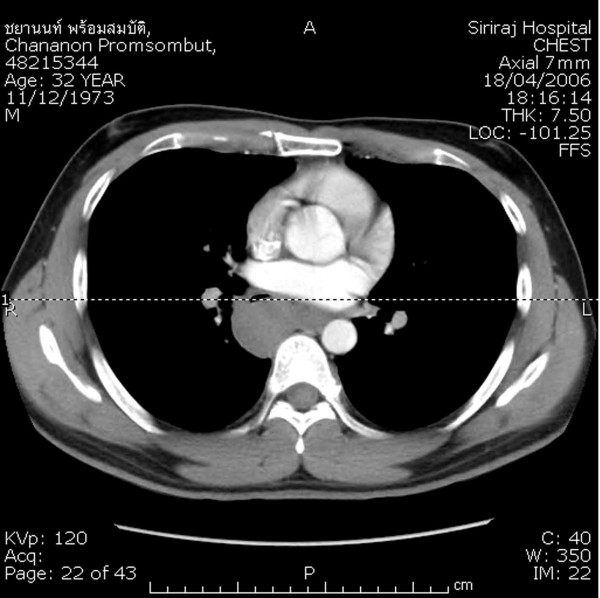
Computed tomography scan of the chest showing the esophageal tumor mass bulging toward the right pleural cavity.
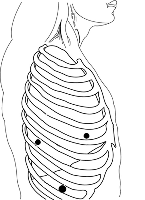
Patient positioning and port sites (A, B and C) for the right side. A: 5-mm port, posterior axillary line, seventh intercostal space. B: 5-mm port, anterior axillary line, fifth intercostal space. C: Camera port, mid-axillary line, nineth intercostal space.
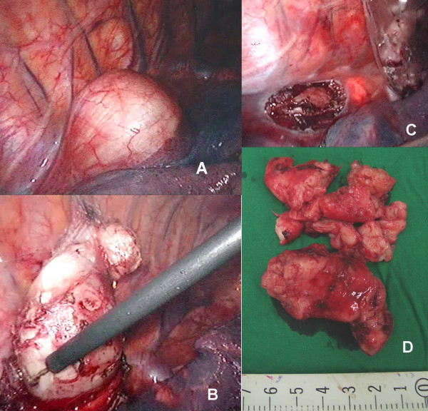
Thoracoscopic findings: (A) The esophageal tumor projects into the right thoracic space; (B) The tumor is enucleated with a simple hook-electrocautery; (C) Trans-illumination from intraopeative esophagoscopy was identified after the tumor was collected in a plastic bag; (D) The tumor was completely removed through camera port in small pieces.
Similar articles
-
Thoracoscopic enucleation of esophageal submucosal tumor by prone position under artificial pneumothorax by CO2 insufflation.Surg Laparosc Endosc Percutan Tech. 2014 Apr;24(2):e55-8. doi: 10.1097/SLE.0b013e31828f71e3. Surg Laparosc Endosc Percutan Tech. 2014. PMID: 24686363
-
Thoracoscopic enucleation of an esophageal leiomyoma.J Clin Gastroenterol. 2000 Jul;31(1):89-90. doi: 10.1097/00004836-200007000-00023. J Clin Gastroenterol. 2000. PMID: 10914787
-
Intraoperative esophagoscopy provides accuracy and safety in video-assisted thoracoscopic enucleation of benign esophageal submucosal tumors.Dis Esophagus. 2015 Jul;28(5):437-41. doi: 10.1111/dote.12220. Epub 2014 Apr 9. Dis Esophagus. 2015. PMID: 24712727
-
[Thoracoscopic enucleation of leiomyoma of the esophagus--report of two cases].Nihon Kyobu Geka Gakkai Zasshi. 1995 Feb;43(2):216-20. Nihon Kyobu Geka Gakkai Zasshi. 1995. PMID: 7714387 Review. Japanese.
-
Thoracoscopic resection of a giant leiomyoma of the esophagus with a mediastinal outgrowth.Ann Thorac Cardiovasc Surg. 1998 Dec;4(6):351-3. Ann Thorac Cardiovasc Surg. 1998. PMID: 9914465 Review.
Cited by
-
Thoracoscopic enucleation of a large esophageal leiomyoma in the lower esophagus: challenges and solutions.Indian J Thorac Cardiovasc Surg. 2021 Nov;37(6):694-697. doi: 10.1007/s12055-021-01196-z. Epub 2021 May 26. Indian J Thorac Cardiovasc Surg. 2021. PMID: 34776669 Free PMC article.
-
Robot-assisted thoracoscopic enucleation for a large esophageal leiomyoma: a case report.Surg Case Rep. 2021 May 26;7(1):129. doi: 10.1186/s40792-021-01212-9. Surg Case Rep. 2021. PMID: 34037886 Free PMC article.
-
Thoracoscopic enucleation of esophageal leiomyoma in prone position and single lumen endotracheal intubation.Surg Endosc. 2013 Sep;27(9):3364-9. doi: 10.1007/s00464-013-2918-3. Epub 2013 Apr 3. Surg Endosc. 2013. PMID: 23549763
-
Prone Position Is Useful in Thoracoscopic Enucleation of Esophageal Leiomyoma.Case Rep Gastroenterol. 2015 May 22;9(2):165-70. doi: 10.1159/000382071. eCollection 2015 May-Aug. Case Rep Gastroenterol. 2015. PMID: 26120297 Free PMC article.
-
Enhanced Wound Healing after Leiomyoma Enucleation.World J Plast Surg. 2018 Jan;7(1):122-127. World J Plast Surg. 2018. PMID: 29651403 Free PMC article.
References
-
- Everitt NJ, Glinatsis M, McMahon MJ. Thoracoscopic enucleation of leiomyoma of the esophagus. Br J Surg. 1992;79:643. - PubMed
LinkOut - more resources
Full Text Sources
