. 2007 Jan;25(1):51-61.
doi: 10.1002/jor.20264.

Ischemia leads to delayed union during fracture healing: a mouse model

Affiliations

Ischemia leads to delayed union during fracture healing: a mouse model

Chuanyong Lu et al. J Orthop Res. 2007 Jan.

Abstract

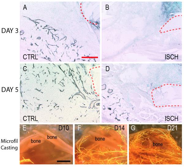
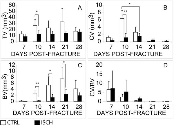
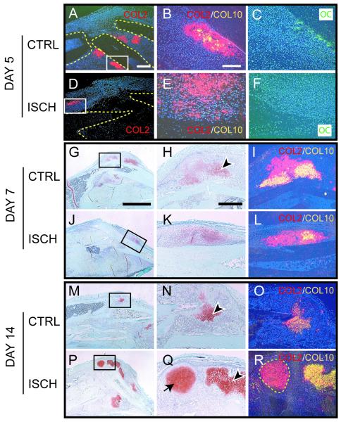
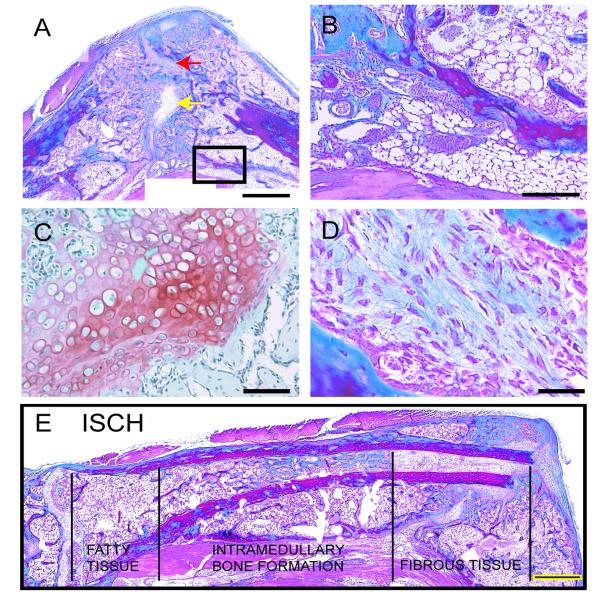
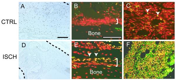
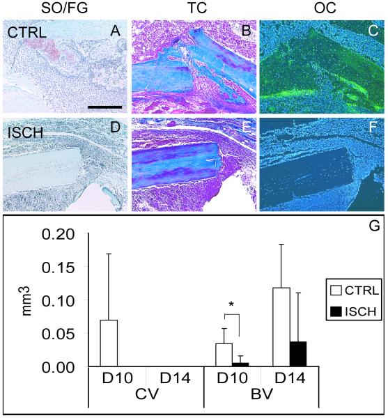
Vascular damage accompanying skeletal injury leads to an ischemic environment, and in clinical settings the extent of vascular damage is directly correlated with failure of skeletal repair. However, the exact mechanism(s) underlying ischemia-related defects in bone healing are not well understood. To better understand the mechanism and to facilitate development of novel interventions to treat ischemic fractures, a mouse model of long bone fracture healing in an ischemic environment was created. Ischemia was induced by femoral artery resection prior to tibia fracture. Fractures were left unstabilized or were stabilized with custom-designed external fixators. Animals with intact femoral vessels served as controls. Tissues from non-stabilized fractures were analyzed at various times from 3 to 28 days after injury (n = 5/time point). Femoral artery resection severely impaired blood supply to the fractured limbs, and perfusion to the fracture sites did not recover until 14 days post-injury. Ischemia significantly decreased the callus size (p < 0.05), and decreased bone (p < 0.05) and cartilage (p < 0.05) matrix production during healing of non-stabilized fracture. The decreased formation of skeletal tissues in ischemic limbs was accompanied by decreased cell proliferation and increased apoptosis at early time points, and increased fibrous and fatty tissues adjacent to the fracture site during the third and fourth week after injury. These alterations led to a delayed-union. Complete fracture healing was not achieved in the majority (day 21 = 4/5; day 28 = 5/5) of ischemic animals, while all control mice (n = 5/5) had evidence of bony bridging by day 21. The ratio of cartilage to bone was similar in ischemic and control limbs at days 7 and 10 in non-stabilized fractures. In stabilized fractures, which healed through direct bone formation in the nonischemic controls, ischemia decreased the amount of bone formation at days 10 and 14 (n = 5/time point) but did not induce cartilage formation. These data reveal that an ischemic insult in the hind limb prior to fracture leads to a delayed union or a nonunion, but does not favor formation of cartilage over bone. This model will be useful for testing novel therapeutic regimens to stimulate fracture healing.


Figures

Figure 1
Figure 1. Perfusion of the lower limb gradually returns in ischemic animals
(A) Immunohistochemical detection of PECAM (black) at day 3 post-injury in the fracture callus illustrates the presence of a vascular network near the fracture site in control animals. (B) Little PECAM staining was evident adjacent to the fracture site in ischemic mice at this time. (C) By 5 days after injury a large number of PECAM-positive profiles are present in the fracture callus of control animals. (D) In ischemic animals, blood vessels are visualized adjacent to but not in the fracture callus. (E) Microfil injection at 10 days after fracture illustrates little perfusion to the limb of ischemic fractures. (F) By day 14, and (G) day 21, more perfusion of the lower leg is evident. Dotted red line=fracture ends. CTRL=control. ISCH=ischemic. Scale bar A-D=200μm, E-G =2mm.
Figure 2
Figure 2. Ischemia reduces formation of skeletal tissues during the healing of non-stabilized fractures
(A) Total volume of the callus (TV). (B) Total volume of the cartilage (CV). (C) Total volume of bone (BV). (D) Ratio of cartilage to bone (CV/BV). CTRL=control. ISCH=ischemic. Data shown are mean ± SD. * p<0.05, ** p<0.01.
Figure 3
Figure 3. Differentiation of chondrocytes and osteoblasts and maturation of chondrocytes are delayed in Ischemic Limbs
(A) Col2 transcripts (red) are present in cells adjacent to the fracture ends (outlined) in control limbs at day 5 post-injury. Boxed area is shown in B and C. (B) A superimposition of adjacent in situ hybridization images reveals that some Col2 positive cells (red) are expressing Col10 transcripts (yellow). (C) Osteoblasts are evident and express oc transcripts (green) in the periosteum. (D) A small domain of Col2 expression (red) is observed at a distance from the fracture site (outlined) in ischemic limbs at 5 days post-injury. The boxed area is shown in E and F. (E) Superimposition of Col2 (red) and Col10 (yellow) in situ hybridization images reveals that no mature chondrocytes are present at this time. (F) Lack of oc expression indicates that no new bone is formed in the periosteum at day 5. (G) Low magnification of control fracture callus 7 days after injury stained with Safranin-O/Fast Green (SO/FG). Cartilage matrix is red. (H) High magnification of box in G illustrates that some chondrocytes are hypertrophic (arrowhead). (I) In situ hybridization reveals that a lot of chondrocytes are expressing Col2 (red) and Col10 (yellow). (J) Low magnification of ischemic fracture callus stained with SO/FG shows the location of cartilage (red). (K) High magnification of boxed area in J reveals that few chondrocytes are hypertrophic, and (L) superimposition of Col2 (red) and Col10 (yellow) in situ hybridization images indicate fewer mature chondrocytes at day 7 post-injury. (M) By day 14, a small amount of cartilage remains in control fractures. (N) High magnification of boxed area in M shows nearly all chondrocytes (arrowhead) exhibit a hypertrophic appearance. (O) In situ hybridization reveals that the chondrocytes are co-expressing Col2 (red) and Col10 (yellow). (P) A large amount of cartilage is present in ischemic fractures at day 14. (Q) High magnification of boxed area in P shows both hypertrophic (arrowhead) and immature (arrow) chondrocytes are present in the fracture site of ischemic limbs. (R) Chondrocytes expressing only Col2 (red, outlined) and chondrocytes expressing Col2 (red) and Col10 (yellow) are present at this time. CTRL=control. ISCH=ischemic. Scale bars: A, D=0.5mm; B, C, E, F=200μm; G, J, M, P=2.5mm; H, I, K, L, N, O, Q, R =500μm.
Figure 4
Figure 4. Adipose and Fibrous Tissue Infiltrate the Fracture Site of Ischemic Limbs at Day 28
(A) Trichrome staining of ischemic fracture callus reveals the presence of cartilage (yellow arrow, shown in C) and fibrous tissues (red arrow, shown in D) in the fracture gap at day 28 post-injury. Boxed area is shown in B. (B) Fatty tissues (arrowheads) are present in areas of the injured leg. (C) Safranin-O/Fast Green staining demonstrates the presence of hypertrophic chondrocytes (red) in the fracture gap of ischemic animals. (D) Trichrome staining of fibrous tissue in the fracture gap. (E) A composite image of the injured tibia 28 days after injury illustrates heterogeneity in the composition of the bone marrow cavity. Near the knee joint the marrow cavity is filled with fatty tissues. Bone is evident in the marrow cavity more distally, and fibrous tissue comprises the marrow cavity adjacent to the fracture site. CTRL=control. ISCH=ischemic. Scale bar A, G=1mm; B=500μm; C=100μm; D=50μm.
Figure 5
Figure 5. Ischemia Reduces Cell Proliferation and Increases Cell Death
(A) Immunohistochemical detection of BrdU incorporation in control animals at 3 days after fracture. (B) The periosteum (bracket) exhibits no signs of cell death, but (C) a few nuclei in the bone marrow cavity are TUNEL positive (arrowheads, yellow) in control animals. Nuclei (red) are counterstained with propidium iodide. (D) Immunohistochemistry identifies few cells that have incorporated BrDU in ischemic limbs. (E) Dying cells (arrowheads, yellow) are present in the periosteum (bracket) of the fractured bone and the adjacent mesenchyme of ischemic limbs. (F) In the bone marrow a large amount of cell death is evident (yellow). Black dotted lines: fractured bone. CTRL=control. ISCH=ischemic. Scale bar=200μm.
Figure 6
Figure 6. Ischemia does not Alter the Mode of Healing in Stabilized Fractures
(A) Safranin-O/Fast Green (SO/FG) staining of control, stabilized fractures reveals a small cartilage island (red) 10 days after fracture, and (B) Trichrome staining (TC) illustrates new bone formation. (C) New bone is confirmed by the presence of osteocalcin (oc, green). (D) In ischemic limbs, there is no evidence of cartilage after SO/FG staining. (E) Likewise, no new bone is observed by TC staining. (F) In situ hybridization reveals no oc transcripts. (G) Histomorphometric analysis illustrates the volume of cartilage (CV) and bone (BV) present at days 10 and 14. At day 10 control animals have significantly more bone than ischemic animals. CTRL=control. ISCH=ischemic. Scale bar=500μm. * p<0.05.

References

    1. Bassett CA, Herrmann I. Influence of oxygen concentration and mechanical factors on differentiation of connective tissues in vitro. Nature. 1961;190:460–1. - PubMed
    1. Choi P, Ogilvie C, Thompson Z, et al. Cellular and molecular characterization of a murine non-union model. J Orthop Res. 2004;22:1100–7. - PubMed
    1. Colnot C, Thompson Z, Miclau T, et al. Altered fracture repair in the absence of MMP9. Development. 2003;130:4123–4133. - PMC - PubMed
    1. Dickson KF, Katzman S, Paiement G. The importance of the blood supply in the healing of tibial fractures. Contemp Orthop. 1995;30:489–93. - PubMed
    1. Domm C, Schunke M, Christesen K, et al. Redifferentiation of dedifferentiated bovine articular chondrocytes in alginate culture under low oxygen tension. Osteo Cart. 2002;10:13–22. - PubMed

Publication types

LinkOut - more resources