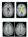
In vivo characterization of traumatic brain injury neuropathology with structural and functional neuroimaging
- PMID: 17020478
- PMCID: PMC1942076
- DOI: 10.1089/neu.2006.23.1396
In vivo characterization of traumatic brain injury neuropathology with structural and functional neuroimaging
Abstract
Quantitative neuroimaging is increasingly used to study the effects of traumatic brain injury (TBI) on brain structure and function. This paper reviews quantitative structural and functional neuroimaging studies of patients with TBI, with an emphasis on the effects of diffuse axonal injury (DAI), the primary neuropathology in TBI. Quantitative structural neuroimaging has evolved from simple planometric measurements through targeted region-of-interest analyses to whole-brain analysis of quantified tissue compartments. Recent studies converge to indicate widespread volume loss of both gray and white matter in patients with moderate-to-severe TBI. These changes can be documented even when patients with focal lesions are excluded. Broadly speaking, performance on standard neuropsychological tests of speeded information processing are related to these changes, but demonstration of specific brain-behavior relationships requires more refined experimental behavioral measures. The functional consequences of these structural changes can be imaged with activation functional neuroimaging. Although this line of research is at an early stage, results indicate that TBI causes a more widely dispersed activation in frontal and posterior cortices. Further progress in analysis of the consequences of TBI on neural structure and function will require control of variability in neuropathology and behavior.
Figures
Similar articles
-
Augmented neural activity during executive control processing following diffuse axonal injury.Neurology. 2008 Sep 9;71(11):812-8. doi: 10.1212/01.wnl.0000325640.18235.1c. Neurology. 2008. PMID: 18779509 Free PMC article.
-
Neuropathology of Mild Traumatic Brain Injury: Correlation to Neurocognitive and Neurobehavioral Findings.In: Kobeissy FH, editor. Brain Neurotrauma: Molecular, Neuropsychological, and Rehabilitation Aspects. Boca Raton (FL): CRC Press/Taylor & Francis; 2015. Chapter 31. In: Kobeissy FH, editor. Brain Neurotrauma: Molecular, Neuropsychological, and Rehabilitation Aspects. Boca Raton (FL): CRC Press/Taylor & Francis; 2015. Chapter 31. PMID: 26269912 Free Books & Documents. Review.
-
The Toronto traumatic brain injury study: injury severity and quantified MRI.Neurology. 2008 Mar 4;70(10):771-8. doi: 10.1212/01.wnl.0000304108.32283.aa. Neurology. 2008. PMID: 18316688
-
Exploring Serum Biomarkers for Mild Traumatic Brain Injury.In: Kobeissy FH, editor. Brain Neurotrauma: Molecular, Neuropsychological, and Rehabilitation Aspects. Boca Raton (FL): CRC Press/Taylor & Francis; 2015. Chapter 22. In: Kobeissy FH, editor. Brain Neurotrauma: Molecular, Neuropsychological, and Rehabilitation Aspects. Boca Raton (FL): CRC Press/Taylor & Francis; 2015. Chapter 22. PMID: 26269900 Free Books & Documents. Review.
-
Neuroimaging and neuropathology of TBI.NeuroRehabilitation. 2011;28(2):63-74. doi: 10.3233/NRE-2011-0633. NeuroRehabilitation. 2011. PMID: 21447905 Review.
Cited by
-
Cerebral atrophy after traumatic white matter injury: correlation with acute neuroimaging and outcome.J Neurotrauma. 2008 Dec;25(12):1433-40. doi: 10.1089/neu.2008.0683. J Neurotrauma. 2008. PMID: 19072588 Free PMC article.
-
Working Memory Deficits After Lesions Involving the Supplementary Motor Area.Front Psychol. 2018 May 23;9:765. doi: 10.3389/fpsyg.2018.00765. eCollection 2018. Front Psychol. 2018. PMID: 29875717 Free PMC article.
-
Multimodal surface-based morphometry reveals diffuse cortical atrophy in traumatic brain injury.BMC Med Imaging. 2009 Dec 31;9:20. doi: 10.1186/1471-2342-9-20. BMC Med Imaging. 2009. PMID: 20043859 Free PMC article.
-
Augmented neural activity during executive control processing following diffuse axonal injury.Neurology. 2008 Sep 9;71(11):812-8. doi: 10.1212/01.wnl.0000325640.18235.1c. Neurology. 2008. PMID: 18779509 Free PMC article.
-
Cognitive processing speed and the structure of white matter pathways: convergent evidence from normal variation and lesion studies.Neuroimage. 2008 Aug 15;42(2):1032-44. doi: 10.1016/j.neuroimage.2008.03.057. Epub 2008 Apr 11. Neuroimage. 2008. PMID: 18602840 Free PMC article.
References
-
- ABDEL-DAYEM HM, ABU-JUDEH H, KUMAR M, et al. SPECT brain perfusion abnormalities in mild or moderate traumatic brain injury. Clin Nucl Med. 1998;23:309–317. - PubMed
-
- ADAMS JH, DOYLE D, FORD I, GENNARELLI TA, GRAHAM DI, MCLELLAN DR. Diffuse axonal injury in head injury: definition, diagnosis and grading. Histopathology. 1989;15:49–59. - PubMed
-
- ADAMS JH, GRAHAM DI, MURRAY LS, SCOTT G. Diffuse axonal injury due to nonmissile head injury in humans: an analysis of 45 cases. Ann Neurol. 1982;12:557–563. - PubMed
-
- ALAVI A, MIROT A, NEWBERG A, et al. Fluorine-18-FDG evaluation of crossed cerebellar diaschisis in head injury. J Nucl Med. 1997;38:1717–1720. - PubMed
-
- ANDERSON CV, BIGLER ED. The role of caudate nucleus and corpus callosum atrophy in trauma-induced anterior horn dilation. Brain Inj. 1994;8:565–569. - PubMed
Publication types
MeSH terms
Grants and funding
LinkOut - more resources
Full Text Sources


