Review
. 2006;7(9):236.
doi: 10.1186/gb-2006-7-9-236.

Arrestins: ubiquitous regulators of cellular signaling pathways

Affiliations
Review

Arrestins: ubiquitous regulators of cellular signaling pathways

Eugenia V Gurevich et al. Genome Biol. 2006.

Abstract

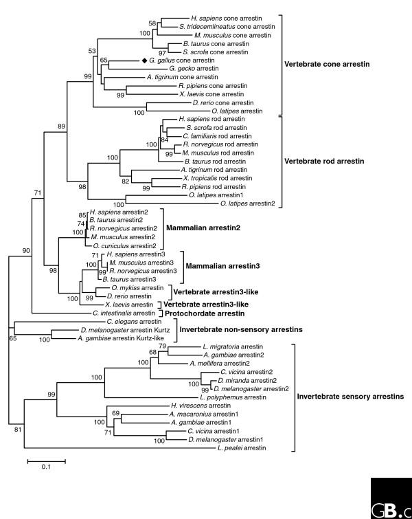
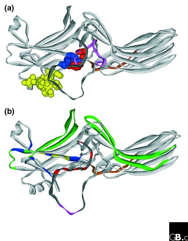
In vertebrates, the arrestins are a family of four proteins that regulate the signaling and trafficking of hundreds of different G-protein-coupled receptors (GPCRs). Arrestin homologs are also found in insects, protochordates and nematodes. Fungi and protists have related proteins but do not have true arrestins. Structural information is available only for free (unbound) vertebrate arrestins, and shows that the conserved overall fold is elongated and composed of two domains, with the core of each domain consisting of a seven-stranded beta-sandwich. Two main intramolecular interactions keep the two domains in the correct relative orientation, but both of these interactions are destabilized in the process of receptor binding, suggesting that the conformation of bound arrestin is quite different. As well as binding to hundreds of GPCR subtypes, arrestins interact with other classes of membrane receptors and more than 20 surprisingly diverse types of soluble signaling protein. Arrestins thus serve as ubiquitous signaling regulators in the cytoplasm and nucleus.


Figures

Figure 1
Figure 1
A phylogenetic tree of the arrestin family. Amino-acid sequence alignments were performed using ClustalW in the MEGA3 software. The phylogenetic tree was created using the neighbor-joining method (gap settings: pairwise deletions; distance method: number of differences). Numbers at selected nodes indicate the percentage frequencies of branch associations on the basis of 1,000 bootstrap repetitions (all percentages over 50 are displayed). Brackets on the right indicate subfamilies. The proteins included for each species, with accession numbers, are as follows: Ambystoma tigrinum (tiger salamander) rod arrestin (AAF14636) and cone arrestin (AAF14637); Anopheles gambiae (African malaria mosquito) arrestin1 (AAG54081), arrestin2 (DAA00888) and Kurtz-like (DAA00889); Apis mellifera (honey bee) XP_623243 (predicted); Ascalaphus macaronius (neuropteran insect) CAC36938; Bos taurus (cattle) rod arrestin (NP_851343), cone arrestin (BAA94344), arrestin2 (NP_776668) and arrestin3 (P32120): C. elegans (nematode) NP_508183; Calliphora vicina (bluebottle fly) arrestin1 (P51486) and arrestin2 (P51487); Canis familiaris (dog) rod arrestin (NP_001003230); C. intestinalis (sea squirt) BAB60819; Danio rerio (zebrafish) cone arrestin (NP_001002405) and arrestin3 (NP_957418); D. melanogaster (fruit fly) arrestin1 (NP_476681), arrestin2 (NP_523976) and Kurtz (NP_524988); Drosophila miranda (fruit fly) arrestin2 (P19108); Gallus gallus (chicken) cone arrestin (XP_420156, predicted); Gekko gecko (tokay) cone arrestin (AAQ94621); Heliothis virescens (tobacco budworm) AAB25861; Homo sapiens (human) rod arrestin (NP_000532), cone arrestin (AAB84302), arrestin2 (NP_004032 isoform A) and arrestin3 (NP_004304 isoform 1); Limulus polyphemus (Atlantic horseshoe crab) P51484; Locusta migratoria (migratory locust, insect) P32122; Loligo pealei (squid) AAK84368; Mus musculus (mouse) rod arrestin (AAH16498), cone arrestin (AAG38954), arrestin2 (NP_796205 isoform A) and arrestin3 (NP_663404); Oncorhynchus mykiss (rainbow trout) arrestin (P51466); Oryctolagus cuniculus (rabbit) arrestin2 (AAC48753); Oryzias latipes (killifish) rod1 arrestin (BAA82259), rod2 arrestin (BAA21718) and cone arrestin (BAA21719); Rana pipiens (northern leopard frog) rod (CAA63135) and cone (CAA63137); Rattus norvegicus (rat) rod (NP_037155), arrestin2 (NP_037042) and arrestin3 (NP_037043); Spermophilus tridecemlineatus (squirrel) cone arrestin (AAS89816); Sus scrofa (pig) rod arrestin (NP_999244) and cone arrestin (NP_999510); Xenopus laevis (frog) cone arrestin (AAH94203) and arrestin3-like (AAH76815); and Xenopus tropicalis rod arrestin (NP_989073).
Figure 2
Figure 2
Key structural elements of arrestin proteins. This model of a generic arrestin molecule was generated in ViewerPro using the crystal structures of bovine rod arrestin [8] and arrestin2 [7]. The proximal carboxy-terminal tail (dark gray) missing in the structures has been modeled arbitrarily. (a) Intra-molecular interactions holding arrestin in the basal conformation. The structure is shown in ribbon representation, except for the residues in the polar core (blue, positive charges; red, negative charges) and the hydrophobic residues in the three-element interaction (yellow), which are shown in space-filling representation. Dark gray indicates the carboxy-terminal tail; magenta, the lariat loop in the carboxy-terminal domain containing two polar core negative charges; light brown, the inter-domain hinge (at the back of the molecule). The polar core is a cluster of five virtually solvent-excluded charged residues, which is unusual for a soluble protein; it includes one negative and one positive charge in the amino-terminal domain, two negative charges in the lariat loop of the carboxy-terminal domain and one positive charge in the carboxy-terminal tail. The three-element interaction is mediated by clusters of bulky hydrophobic residues in β-strand I, α-helix I and the carboxy-terminal tail. (b) Known interaction sites on the arrestin molecule. Receptor-binding elements: blue, positive charges that bind receptor-attached phosphates [70]; yellow, hydrophobic residues in β-strand X [71]; green, elements that determine receptor specificity [30]. Other elements: magenta, the clathrin-binding element in the proximal carboxy-terminal tail [27]; red, AP2-binding residues in the distal carboxy-terminal tail [28].

Similar articles

Cited by

References

    1. Gurevich VV, Gurevich EV. The structural basis of arrestin-mediated regulation of G-protein-coupled receptors. Pharmacol Ther. 2006;110:465–502. doi: 10.1016/j.pharmthera.2005.09.008. - DOI - PMC - PubMed
    1. Sakuma H, Murakami A, Fujimaki T, Inana G. Isolation and characterization of the human X-arrestin gene. Gene. 1998;224:87–95. doi: 10.1016/S0378-1119(98)00510-1. - DOI - PubMed
    1. Yamaki K, Tsuda M, Kikuchi T, Chen KH, Huang KP, Shinohara T. Structural organization of the human S-antigen gene. cDNA, amino acid, intron, exon, promoter, in vitro transcription, retina, and pineal gland. J Biol Chem. 1990;265:20757–20762. - PubMed
    1. Nakagawa M, Orii H, Yoshida N, Jojima E, Horie T, Yoshida R, Haga T, Tsuda M. Ascidian arrestin (Ci-arr), the origin of the visual and nonvisual arrestins of vertebrate. Eur J Biochem. 2002;269:5112–5118. doi: 10.1046/j.1432-1033.2002.03240.x. - DOI - PubMed
    1. Palmitessa A, Hess HA, Bany IA, Kim YM, Koelle MR, Benovic JL. Caenorhabditis elegans arrestin regulates neural G protein signaling and olfactory adaptation and recovery. J Biol Chem. 2005;280:24649–24662. doi: 10.1074/jbc.M502637200. - DOI - PubMed

Publication types