

Observations on the management of Coats' disease: less is more
- PMID: 17020897
- PMCID: PMC1857684
- DOI: 10.1136/bjo.2006.103382
Observations on the management of Coats' disease: less is more
Abstract
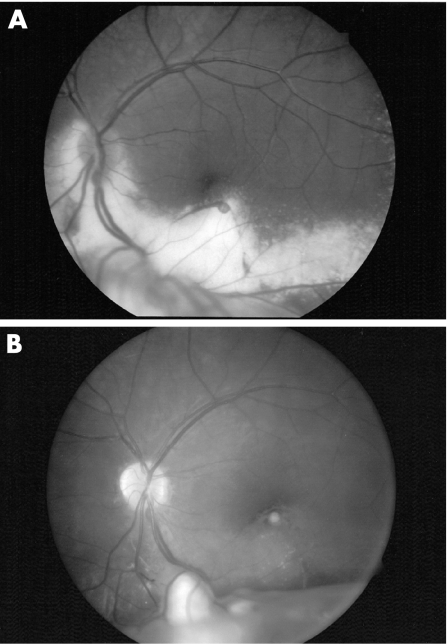
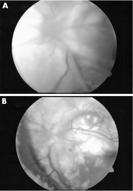
Background: In this article we share our experience of treating various severities of Coats' disease and focus on optimal therapy for advanced disease.
Methods: Retrospective chart review of 10 patients treated with varied techniques including intraocular surgery, cryopexy and/or laser photocoagulation.
Results: Nine patients were male. At presentation the average age was 4.6 years (range 21 months-7 years), the average number of retinal quadrants involved with telangiectasia was 2.7 (range 1-4, median 3), eight of the 10 patients had retinal detachment, six of these being total, and all patients had macular involvement with either exudate or fibrosis. Average follow-up was 2.3 years (range 1-4.5 years). The best visual outcomes were observed in patients who presented with less severe disease. For example, the only four patients to maintain ambulatory vision all presented without total retinal detachment, two or fewer quadrants of retinal telangiectasia and a visual acuity better than light perception. No patient developed secondary angle closure glaucoma, and all patients have kept a cosmetically acceptable eye.
Conclusion: In this limited series, visual outcomes in the setting of advanced Coats' disease are largely dependent on disease severity and visual status at the time of presentation. Minimally invasive surgery with vitreous infusion through the pars plana, combined with external drainage of subretinal fluid together with cryotherapy and/or laser photocoagulation is sufficient to effect retinal re-attachment and prevent loss of the eye.
Conflict of interest statement
Competing interests: None of the authors have a proprietary interest in this work.
References
-
- Coats G. Forms of retinal disease with massive exudation. Roy London Ophthal Hosp Rep 190817440–525.
-
- Harris G S. Coats' disease, diagnosis and treatment. Mod Probl Ophthal 197210277–285. - PubMed
-
- Harris G S. Coats' disease, diagnosis and treatment. Can J Ophthalmol 19705311–319. - PubMed
-
- Deutsch T A, Robb M F, Jampol L M. Spontaneous regression of retinal lesions in Coats' disease. Can J Ophthalmol 198217169–172. - PubMed
-
- Morales A G. Coats' disease: natural history and results of treatment. Am J Ophthalmol 196560855–865. - PubMed
MeSH terms
LinkOut - more resources
Full Text Sources
Medical
Miscellaneous