Comparative Study
. 2006 Dec;80(24):11935-45.
doi: 10.1128/JVI.00621-06. Epub 2006 Oct 4.

Analysis of the cytosolic domains of the hepatitis B virus envelope proteins for their function in viral particle assembly and infectivity

Affiliations
Comparative Study

Analysis of the cytosolic domains of the hepatitis B virus envelope proteins for their function in viral particle assembly and infectivity

Matthieu Blanchet et al. J Virol. 2006 Dec.

Abstract

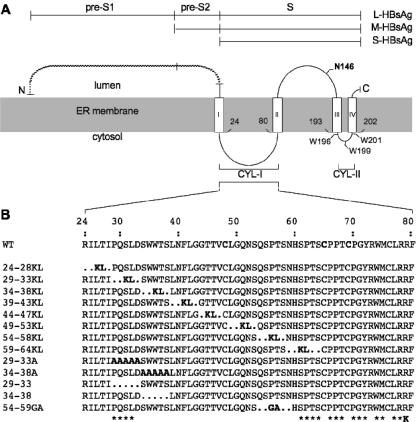
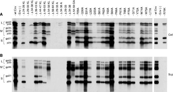
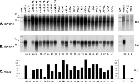
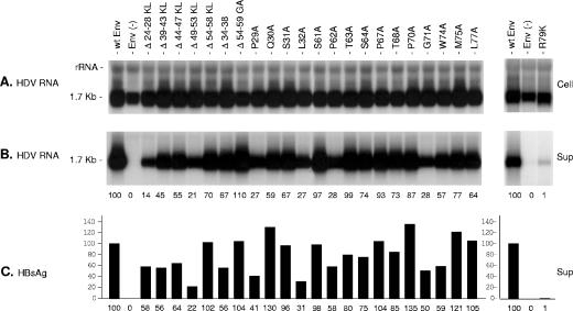
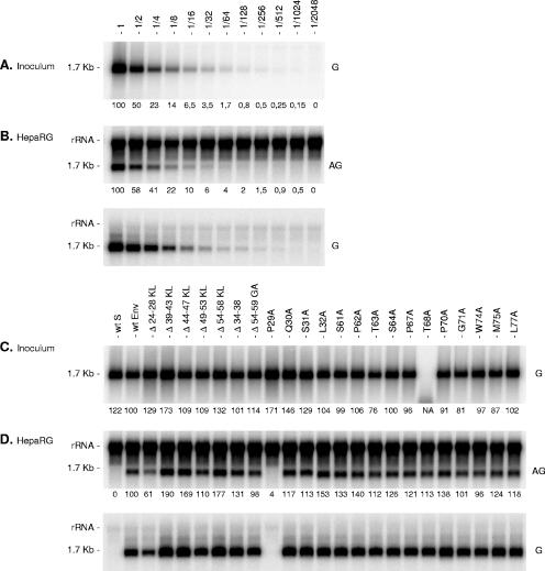
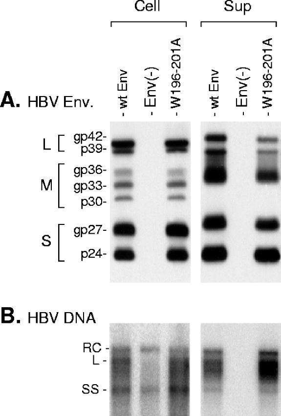
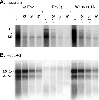
The hepatitis B virus (HBV) envelope proteins have the ability to assemble three types of viral particles, (i) the empty subviral particles (SVPs), (ii) the mature HBV virions, and (iii) the hepatitis delta virus (HDV) particles, in cells that are coinfected with HBV and HDV. To gain insight into the function of the HBV envelope proteins in morphogenesis of HBV or HDV virions, we have investigated subdomains of the envelope proteins that have been shown or predicted to lie at the cytosolic face of the endoplasmic reticulum membrane during synthesis, a position prone to interaction with the inner core structure. These domains, referred to here as cytosolic loops I and II (CYL-I and -II, respectively), were subjected to mutagenesis. The mutations were introduced in the three HBV envelope proteins, designated small, middle, and large (S-HBsAg, M-HBsAg, and L-HBsAg, respectively). The mutants were expressed in HuH-7 cells to evaluate their capacity for self-assembly and formation of HBV or HDV virions when HBV nucleocapsid or HDV ribonucleoprotein, respectively, was provided. We found that SVP-competent CYL-I mutations between positions 23 and 78 of the S domain were permissive to HBV or HDV virion assembly. One mutation (P29A) was permissive for synthesis of the S- and M-HBsAg but adversely affected the synthesis or stability of L-HBsAg, thereby preventing the assembly of HBV virions. Furthermore, using an in vitro infection assay based on the HepaRG cells and the HDV model, we have shown that particles coated with envelope proteins bearing CYL-I mutations were fully infectious, hence indicating the absence of an infectivity determinant in this region. Finally, we demonstrated that the tryptophan residues at positions 196, 199, and 201 in CYL-II, which were shown to exert a matrix function for assembly of HDV particles (I. Komla-Soukha and C. Sureau, J. Virol. 80:4648-4655, 2006), were dispensable for both assembly and infectivity of HBV virions.


Figures

FIG. 1.
FIG. 1.
Schematic representations of envelope protein mutants. (A) L-, M-, and S-HBsAg proteins are depicted by horizontal lines. The topology of the envelope protein S domains at the ER membrane is represented. Open boxes represent transmembrane regions in the envelope proteins S domain, and the shaded area corresponds to the ER membrane. Experimentally defined (I and II) or putative (III and IV) transmembrane signals are indicated. (B) CYL-I sequences of the envelope protein mutants are indicated. Mutants are designated by the positions of the first and last deleted amino acids followed by the one-letter codes of the inserted residues. Asterisks indicate the positions of amino acids that were substituted with alanine in mutants presenting single amino acid substitutions. The R79K mutation (K) is indicated.
FIG. 2.
FIG. 2.
Effects of CYL-I mutations on envelope protein synthesis and secretion. (A) Cellular proteins harvested at day 4 posttransfection (Cell) were separated on an acrylamide gel, transferred to a PVDF membrane, and probed with a mixture of anti-S and anti-preS2 antibodies (1:500 and 1:1,000, respectively). Signals are from 105 cells. (B) A pool of culture medium collected at days 2 and 4 posttransfection (Sup) was subjected to ultracentrifugation. Sedimented particles were disrupted in Laemmli buffer and analyzed as described above. Signals are from 1 ml of culture medium. Env(−), envelope protein negative; wt SML, wt S-, M-, and L-HBsAg. Molecular weights of the glycosylated (gp) and unglycosylated (p) forms of the HBV envelope proteins are indicated.
FIG. 3.
FIG. 3.
Effects of envelope protein CYL-I mutations on secretion of HBV virions. (A) Southern blot analysis of intracellular HBV DNA. Total DNA from HuH-7 cells (Cell) cotransfected with pCIHBenv(−) and pT7HB2.7 or derivatives was extracted at day 11, separated on a 1% agarose gel, transferred to a nylon membrane, and probed with a 32P-RNA probe specific for negative-strand HBV DNA. Signals are from 105 cells. Molecular weight markers (MW) include 10 pg of cloned linear (L) HBV DNA, corresponding to 3 × 10⁶ ge. (B) Culture media collected at days 3, 6, 9, and 11 posttransfection (Sup) were pooled and subjected to immunoprecipitation using an anti-preS1 antibody. Precipitates were disrupted, and DNA was extracted and analyzed by Southern blot hybridization as described above. Signals are from 1 ml of culture medium. For a positive control (wt Env), the 100% values correspond to 200 pg/ml of HBV DNA (left panel) or 300 pg/ml (right panel), approximately 6 × 10⁷ or 9 × 10⁷ ge/ml, respectively. (C) Histogram showing the relative amounts of secreted HBV envelope proteins measured with an anti-HBsAg ELISA (DiaSorin). For a positive control (wt Env), the 100% value corresponds to approximately 400 ng/ml of HBsAg (left panel) or 150 ng/ml (right panel), equivalent to approximately 1011 or 3.7 × 10¹⁰ SVPs, respectively. RC, relax circular; L, linear; SS, single stranded; Env(−), envelope protein negative; wt SML, wt S-, M-, and L-HBsAg. Numbers below each panel are percentages of the wt value.
FIG. 4.
FIG. 4.
Effects of envelope protein CYL-I mutations on secretion of HDV virions. (A) Northern blot analysis of intracellular HDV RNA. Total RNA from HuH-7 cells (Cell) cotransfected with pSVLD3 and pT7HB2.7 or derivatives was extracted at day 12, separated on a 1.2% agarose gel, transferred to a nylon membrane, and probed with a genomic strand-specific, 32P-labeled RNA probe. Signals were obtained from 105 cells. (B) Northern blot analysis of extra cellular HDV RNA. Culture media collected at days 5, 7, 9, and 12 posttransfection (Sup) were pooled and subjected to ultracentrifugation on a sucrose cushion. RNA was extracted and analyzed as described above. Signals are from 0.5 ml of culture medium. For positive control (wt Env), the 100% values correspond to 800 pg/ml of HDV RNA (left panel) or 400 pg/ml (right panel), which approximate 8 × 10⁸ or 4 × 10⁸ ge, respectively. (C) Histogram showing the relative amounts of secreted HBV envelope proteins measured with an anti-HBsAg ELISA (DiaSorin). For a positive control (wt Env), the 100% value corresponds to approximately 700 ng/ml of HBsAg (left panel) or 300 ng/ml (right panel), equivalent to approximately 1.75 1011 or 7.4 × 10¹⁰ SVPs, respectively. Env(−), envelope protein negative; wt Env, wt S-, M-, and L-HBsAg. Numbers below each panel are percentages of the wt value.
FIG. 5.
FIG. 5.
Effects of envelope protein CYL-I mutations on the infectivity of HDV virions. (A) Supernatant (1 ml) containing approximately 2 × 10⁸ ge of wt HDV virions was serially diluted prior to analysis by Northern blot hybridization with a genomic-strand-specific, 32P-labeled RNA probe (G). One milliliter of undiluted or 2- to 2,048-fold diluted wt HDV-containing medium was added to HepaRG cells in the presence of 4% PEG 8000. (B) Cells were harvested 8 days after inoculation and analyzed for the presence of HDV RNA by Northern blot hybridization with an antigenomic (AG) or a genomic (G) strand-specific, 32P-labeled RNA probe. Dilution factor is indicated. (C) Supernatants (1 ml) from HuH-7 cell cultures were normalized for their genomic HDV RNA concentration. One ml of normalized inoculum was added to HepaRG cells in the presence of 4% PEG 8000. Inocula were recovered after overnight exposure to cells, and their genomic HDV RNA content was controlled by Northern blot hybridization using a genomic strand-specific, 32P-labeled RNA probe. Signals are from 0.5 ml of postexposure inocula. Cells were harvested 8 days after exposure and analyzed for the presence of HDV RNA as described above. rRNA which hybridized nonspecifically to antigenomic-specific HDV RNA probes served as a loading control. The size in kilobases of HDV RNA is indicated. wt S, HDV particles coated with the wt S-HBsAg only. wt Env, HDV particles coated with wt S-, M-, and L-HBsAg. Numbers below each panel are percentages of the wt value. NA, not applicable.
FIG. 6.
FIG. 6.
Effects of CYL-II envelope protein mutations on assembly of HBV virions. Production of HBV particles by transfection of 106 cells with 1 μg of pCIHBenv(−) and 1 μg of pT7HB2.7 (wt Env), an empty plasmid, Env(−), or a W196-201A mutant plasmid. (A) Cellular proteins (Cell) harvested at day 4 posttransfection were separated on an acrylamide gel, transferred to a PVDF membrane, and probed with a mixture of anti-S and anti-preS2 antibodies. Signals are from 105 cells. A pool of culture medium collected at days 3, 6, 9, and 12 posttransfection was subjected to ultracentrifugation. Sedimented particles were disrupted in Laemmli buffer and analyzed as described above. Signals (Sup) are from 1 ml of culture medium. (B) Total DNA from transfected cells (Cell) was extracted, separated on a 1% agarose gel, transferred to a nylon membrane, and probed with a 32P-RNA probe specific for negative-strand HBV DNA. Signals are from 105 cells. Culture medium (Sup) was subjected to immunoprecipitation using an anti-preS1 antibody. Precipitates were disrupted, and DNA was extracted and analyzed by Southern blot hybridization as described above. Signals are from 1 ml of culture medium. The L-HBsAg, M-HBsAg, and S-HBsAg proteins are indicated (L, M, and S respectively). Env(−), envelope proteins negative; wt Env, wt S-, M-, and L-HBsAg. Molecular weights of the glycosylated (gp) and unglycosylated (p) forms of the HBV envelope proteins are indicated. RC, relax circular; L, linear; SS, single stranded.
FIG. 7.
FIG. 7.
Effects of CYL-II envelope protein mutations on infectivity of HBV virions. (A) Culture medium from cells transfected with pCIHBenv(−) and 1 μg of pT7HB2.7 (wt Env), an empty plasmid, Env(−), or W196-201A mutant plasmid, collected at days 3, 6, 9, and 11 posttransfection (Sup), were pooled and subjected to a 20× concentration by precipitation with PEG 8000. One hundred microliters of undiluted (1) or a two- (1/2), four- (1/4), or eightfold (1/8) dilution of each inoculum was examined for the presence of HBV DNA. HBV DNA signals in the inoculum derived from cells transfected in the absence of envelope protein coding plasmid [Env(−)] are from extracellular nonenveloped nucleocapsids, whereas signals from wt Env and W196-201A samples are from both naked nucleocapsids and virions. (B) HepaRG cells (3.3 × 10⁵ cells) were exposed to 2 ml of inoculum for 16 h in the presence of 5% PEG 8000. HepaRG cells were harvested at day 12 postinoculation, and mRNA was purified and tested for the presence of newly synthesized HBV mRNA by Northern blot hybridization using a negative-strand-specific, 32P-labeled RNA probe. RC, relax circular; L, linear; SS, single strand. The sizes in kilobases of HBV mRNAs are indicated.

References

    1. Barrera, A., B. Guerra, L. Notvall, and R. E. Lanford. 2005. Mapping of the hepatitis B virus pre-S1 domain involved in receptor recognition. J. Virol. 79:9786-9798. - PMC - PubMed
    1. Bonino, F., K. H. Heermann, M. Rizzetto, and W. H. Gerlich. 1986. Hepatitis delta virus: protein composition of delta antigen and its hepatitis B virus-derived envelope. J. Virol. 58:945-950. - PMC - PubMed
    1. Bruss, V. 1997. A short linear sequence in the pre-S domain of the large hepatitis B virus envelope protein required for virion formation. J. Virol. 71:9350-9357. - PMC - PubMed
    1. Bruss, V., and D. Ganem. 1991. The role of envelope proteins in hepatitis B virus assembly. Proc. Natl. Acad. Sci. USA 88:1059-1063. - PMC - PubMed
    1. Bruss, V., X. Lu, R. Thomssen, and W. H. Gerlich. 1994. Post-translational alterations in transmembrane topology of the hepatitis B virus large envelope protein. EMBO J. 13:2273-2279. - PMC - PubMed

Publication types

Substances

LinkOut - more resources