Comparative Study
. 2006 Dec;80(24):12109-20.
doi: 10.1128/JVI.01370-06. Epub 2006 Oct 4.

A new group B adenovirus receptor is expressed at high levels on human stem and tumor cells

Affiliations
Comparative Study

A new group B adenovirus receptor is expressed at high levels on human stem and tumor cells

Sebastian Tuve et al. J Virol. 2006 Dec.

Abstract

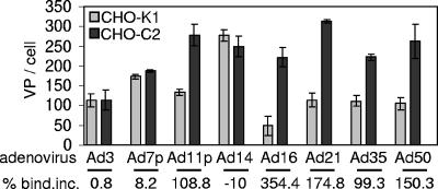
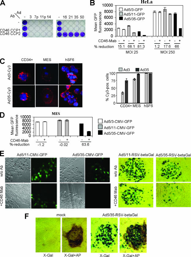
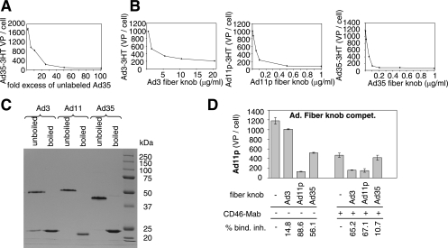
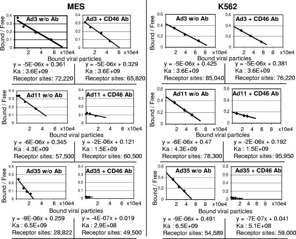
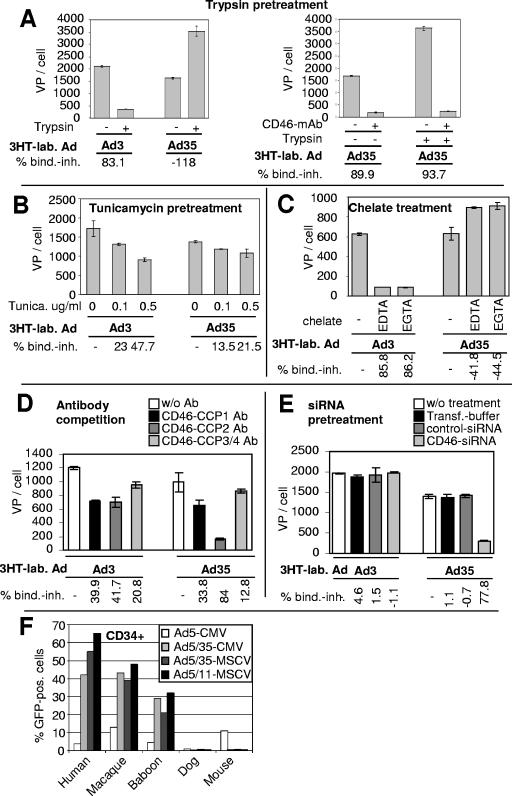
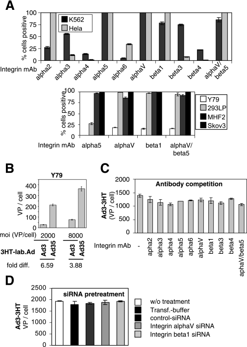
CD46 is used by human group B adenoviruses (Ads) as a high-affinity attachment receptor. Here we show evidence that several group B Ads utilize an additional receptor for infection of human cells, which is different from CD46. We tentatively named this receptor receptor X. Competition studies with unlabeled and labeled Ads, recombinant Ad fiber knobs, and soluble CD46 and CD46 antibodies revealed three different subgroups of group B Ads, in terms of their receptor usage. Group I (Ad16, -21, -35, and -50) nearly exclusively uses CD46. Group II (Ad3, -7p, and -14) utilizes receptor X and not CD46. Group III (Ad11p) uses both CD46 and the alternative receptor X. Interaction of group II and III Ads with receptor X occurs via the fiber knob. Receptor X is an abundantly expressed glycoprotein that interacts with group II and III Ads at relatively low affinity in a Ca(2+)-dependent manner. This receptor is expressed at high levels on human mesenchymal and undifferentiated embryonic stem cells, as well as on human cancer cell lines. These findings have practical implications for stem cell and gene therapy.


Figures

FIG. 1.
FIG. 1.
Attachment study with group B Ads on CHO-K1 versus CHO-C2 cells. 3H-labeled Ads were added, and the number of VP bound per cell was determined after 1 h of incubation on ice.
FIG. 2.
FIG. 2.
Role of CD46 in group B Ad infection. (A) CPE assay with group B Ads on HeLa cells. A total of 2 × 10⁵ HeLa cells/well were seeded in 24-well plates. Twenty-four hours later, cells were preincubated with antibodies directed against the CCP1 or CCP2 domain of CD46 for 30 min (10 μg/ml). Ads were then added at an MOI of 100 PFU/cell (∼2,000 VP/cell). Forty-eight hours after infection, cells were washed with PBS, fixed with 4% paraformaldehyde, and stained with crystal violet as described in Materials and Methods. (B) Fiber chimeric Ad infection blocking study. HeLa cells were preincubated with the MEM-258 MAb and then infected with Ad5/3-CMV-GFP, Ad5/11-CMV-GFP, or Ad5/35-CMV-GFP at an MOI of 25 or 250 PFU/cell, and GFP fluorescence was assessed by flow cytometry 16 h after infection. (C) Ad-Cy3 binding to primary cells. Primary cells were incubated with Ad3-Cy3 or Ad35-Cy3 either in suspension (CD34+, hSF6) or attached to chamber slides (MES). Cell nuclei were counterstained with 4′,6′-diamidino-2-phenylindole (DAPI) (blue). Cy3-labeled VP appear as red dots on the cell surface. Magnifications were ×20 (MES), ×40 (hSF6), and ×100 (CD34+). The graph on the right shows the average percentage of Cy3-positive cells from 10 random viewing fields (magnification, ×40). (D) MES cells were preincubated with the MEM-258 MAb for 30 min at RT (10 μg/ml). Cells were then infected with Ad5/3-CMV-GFP, Ad5/11-CMV-GFP, or Ad5/35-CMV-GFP at an MOI of 25 PFU/cell. GFP fluorescence was assessed by flow cytometry 24 h after infection. (E) hSF6 cells were preincubated with the MEM-258 MAb for 30 min at RT (10 μg/ml). Cells were then infected with Ad5/11-CMV-GFP or Ad5/35-CMV-GFP (left side). Representative samples of GFP fluorescence 24 h after infection and the corresponding cell morphology are shown. w/o Ab, without antibody. (Right side) Infection was performed as described above, with Ad5/11-RSV-β-Gal and Ad5/35-RSV-β-Gal. At 24 h after infection, cells were fixed and stained for β-Gal expression. Magnification, ×20. (F) hSF6 cells were mock infected or infected with Ad5/35-RSV-β-Gal at an MOI of 250 PFU/cell. At 24 h after infection, cells were fixed and stained for β-Gal expression (X-Gal) and then for alkaline phosphatase (X-Gal+AP) as described in Materials and Methods. β-Gal-positive cells show a green-blue nucleus and slightly green cytoplasm. AP-positive cells show brown cytoplasm. Representative samples are shown.
FIG. 3.
FIG. 3.
Group B Ad-cell interaction occurs via fiber knob. (A) Cross competition of Ad35 for attachment to HeLa cells. Cells were incubated with a 2- to 100-fold excess of unlabeled Ad35. Thereafter, 3H-labeled Ad35 was added at an MOI of 8,000 VP per cell and the number of VP bound per cell was determined. (B) Fiber knob competition of Ad3, Ad11p, and Ad35 attachment to HeLa cells. Cells were preincubated with various concentrations of the recombinant fiber knob proteins of Ad3 (1 to 20 μg/ml), Ad11p (0.01 to 1 μg/ml), and Ad35 (0.01 to 1 μg/ml). Corresponding 3H-labeled Ads were then added at an MOI of 8,000 VP per cell, and numbers of VP bound per cell were determined. (C) Analysis of recombinant Ad3, Ad11p, and Ad35 fiber knob proteins by electrophoresis (unboiled versus boiled for 3 min at 100°C) on a 10% polyacrylamide gel. The bands were visualized by brilliant blue R-250 (Fisher Biotech) stain reagent. The values on the right are molecular masses of marker proteins in kilodaltons. (D) Fiber knob competition of Ad11p attachment to HeLa cells. Cells were preincubated with the MEM-258 MAb, and subsequently recombinant fiber knob proteins of Ad3 (10 μg/ml), Ad11p (1 μg/ml), and Ad35 (1 μg/ml) were added. Thereafter, 3H-labeled Ad11p was added at an MOI of 8,000 VP per cell and attachment was analyzed. bind. inh., binding inhibition.
FIG. 4.
FIG. 4.
Affinity and receptor numbers. Ka values and receptor sites for Ad3, -11p, and -35 on MES cells and K562 cells are shown. w/o Ab, without antibody.
FIG. 5.
FIG. 5.
Characterization of receptor X. All data shown were acquired with HeLa cells. (A) Cells were detached from culture dishes with trypsin (+) or EDTA (−). Cells were preincubated with MEM-258 MAb (right side). 3H-labeled (3HT-lab.) Ad3 or Ad35 was then added at an MOI of 8,000 VP per cell. (B) Cells were pretreated with tunicamycin (Tunica.) at the concentrations shown for 48 h. Then, 3H-labeled Ad3 or Ad35 was added at an MOI of 8,000 VP per cell and the numbers of VP per cell were determined. (C) Cells were incubated with 3H-labeled Ad3 or Ad35 (MOI, 8,000 VP/cell) in the presence of 10 mM EDTA or EGTA. Numbers of VP bound per cell were determined. (D) Antibody competition of Ad attachment. Cells were preincubated with CD46-CCP1-, CD46-CCP2-, or CD46-CCP3/4-specific MAbs as described in Materials and Methods. Thereafter 3H-labeled Ad3 or Ad35 (MOI, 8,000 VP/cell) was added and the numbers of VP bound per cell were measured. (E) Cells were mock transfected or transfected with control siRNA or CD46 siRNA as described in Materials and Methods. At 48 h after transfection, 3H-labeled Ad3 or Ad35 (8,000 VP/cell) was added and the numbers of VP bound per cell were determined. (F) Fiber chimeric Ad infection study on CD34+ cells of different species. A total of 105 CD34+ cells were infected with GFP-expressing viruses (Ad5-CMV-GFP, Ad5/35-CMV-GFP, Ad5/11-CMV-GFP, Ad5/11-MSCV-GFP) at an MOI of 25 PFU/cell in suspension in 300 μl. GFP fluorescence intensities were assessed by flow cytometry 24 h after infection. MSCV, murine stem cell virus. w/o Ab, without antibody; bind. inh., binding inhibition.
FIG. 6.
FIG. 6.
Assessment of integrins as receptor X candidates. (A) Flow cytometry analysis of integrin expression on K562, HeLa, Y79, 293, MHF2, and SKOV3 cells was carried out as described in Materials and Methods. Percentages of Alexa Fluor 488-positive cells are shown. (B) Studies of Ad3 and Ad35 attachment to Y79 cells. Y79 cells were incubated with 3H-labeled (3HT-lab.) Ads at an MOI of 2,000 or 8,000 VP per cell, and the numbers of VP bound per cell were determined. diff., difference. (C) Antibody competition of Ad3 binding to HeLa cells. Cells were preincubated with MAbs directed toward integrins (10 μg/ml) as described in Materials and Methods. 3H-labeled Ads (8,000 VP/cell) were then added, and the numbers of VP bound per cell were determined. (D) HeLa cells were mock transfected or transfected with control siRNA or siRNA specific for αv and β1 integrins as described in Materials and Methods. At 48 h after transfection, 3H-labeled Ad3 (8,000 VP/cell) was added and the number of VP bound per cell was determined. Transf., transfer.

References

    1. Anderson, B. D., T. Nakamura, S. J. Russell, and K. W. Peng. 2004. High CD46 receptor density determines preferential killing of tumor cells by oncolytic measles virus. Cancer Res. 64:4919-4926. - PubMed
    1. Barton, E. S., B. E. Youree, D. H. Ebert, J. C. Forrest, J. L. Connolly, T. Valyi-Nagy, K. Washington, J. D. Wetzel, and T. S. Dermody. 2003. Utilization of sialic acid as a coreceptor is required for reovirus-induced biliary disease. J. Clin. Investig. 111:1823-1833. - PMC - PubMed
    1. Bernt, K. M., M. Liang, X. Ye, S. Ni, Z.-Y. Li, S. L. Ye, F. Hu, and A. Lieber. 2002. A new type of adenovirus vector that utilizes homologous recombination to achieve tumor-specific replication. J. Virol. 76:10994-11002. - PMC - PubMed
    1. Czyz, J., and A. Wobus. 2001. Embryonic stem cell differentiation: the role of extracellular factors. Differentiation 68:167-174. - PubMed
    1. DiPaolo, N., S. Ni, A. Gaggar, R. Strauss, S. Tuve, Z. Y. Li, D. Stone, D. Shayakhmetov, N. Kiviat, P. Toure, S. Sow, B. Horvat, and A. Lieber. 2006. Evaluation of adenovirus vectors containing serotype 35 fibers for vaccination. Mol. Ther. 13:756-765. - PMC - PubMed

Publication types

MeSH terms

LinkOut - more resources