Comparative Study
. 2006 Oct 4;26(40):10100-9.
doi: 10.1523/JNEUROSCI.2476-06.2006.

Effects in neocortical neurons of mutations of the Na(v)1.2 Na+ channel causing benign familial neonatal-infantile seizures

Affiliations
Comparative Study

Effects in neocortical neurons of mutations of the Na(v)1.2 Na+ channel causing benign familial neonatal-infantile seizures

Paolo Scalmani et al. J Neurosci. .

Abstract

Mutations of voltage-gated Na+ channels are the most common cause of familial epilepsy. Benign familial neonatal-infantile seizures (BFNIS) is an epileptic trait of the early infancy, and it is the only well characterized epileptic syndrome caused exclusively by mutations of Na(V)1.2 Na+ channels, but no functional studies of BFNIS mutations have been done. The comparative study of the functional effects and the elucidation of the pathogenic mechanisms of epileptogenic mutations is essential for designing targeted and effective therapies. However, the functional properties of Na+ channels and the effects of their mutations are very sensitive to the cell background and thus to the expression system used. We investigated the functional effects of four of the six BFNIS mutations identified (L1330F, L1563V, R223Q, and R1319Q) using as expression system transfected pyramidal and bipolar neocortical neurons in short primary cultures, which have small endogenous Na+ current and thus permit the selective study of transfected channels. The mutation L1330F caused a positive shift of the inactivation curve, and the mutation L1563V caused a negative shift of the activation curve, effects that are consistent with neuronal hyperexcitability. The mutations R223Q and R1319Q mainly caused positive shifts of both activation and inactivation curves, effects that cannot be directly associated with a specific modification of excitability. Using physiological stimuli in voltage-clamp experiments, we showed that these mutations increase both subthreshold and action Na+ currents, consistently with hyperexcitability. Thus, the pathogenic mechanism of BFNIS mutations is neuronal hyperexcitability caused by increased Na+ current.


Figures

Figure 1.
Figure 1.
BNIFS mutations. The position of the six identified BNIFS mutations is indicated on the schematic membrane topology of the Nav1.2 Na+ channel α subunit. The mutation R1319Q has been identified in three different families. The mutations studied in this paper are highlighted by the ovals.
Figure 2.
Figure 2.
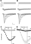
Macropatch Na+ currents recorded in neurons transfected with Nav1.2 Na+ channel α subunit. A, Middle, Macropatch Na+ currents elicited by the voltage stimuli shown above in a representative control bipolar neuron transfected with YFP; bottom, macropatch Na+ currents recorded applying the same voltage stimuli in a representative bipolar neuron transfected with Nav1.2 Na+ channel. Calibration: 50 pA, 1 ms. B, Middle, Macropatch Na+ currents recorded in a representative control pyramidal neuron transfected with YFP and elicited by the voltage stimuli shown above; bottom, macropatch Na+ currents recorded in a representative pyramidal neuron transfected with Nav1.2 and elicited by the voltage stimuli shown in the top. C, Mean macropatch current–voltage curves for control YFP-transfected bipolar neurons (open circles) and bipolar neurons transfected with Nav1.2 (filled circles). D, Mean macropatch current–voltage curves for control YFP-transfected pyramidal neurons (filled pentagons) and pyramidal neurons transfected with Nav1.2 (open triangles).
Figure 3.
Figure 3.
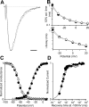
Properties of Nav1.2 Na+ currents in transfected neurons. A, Average normalized macropatch current traces elicited with a depolarizing step to −20 mV for Nav1.2-transfected pyramidal (solid line) and bipolar (dashed line) neurons. Horizontal calibration bar, 500 μs. B, Top, Semilogarithmic plot of the time to half-activation at the indicated potentials for pyramidal (open triangles) and bipolar (filled circles) neurons; bottom, time constant of inactivation derived from fits of exponential functions to the decay of the current traces during depolarizations to the indicated potentials. C, Mean voltage dependence of activation and inactivation; the lines are Boltzmann relationships whose parameters were calculated averaging the parameters of the fits of the single cells (Table 1). D, Mean kinetics of recovery from a 100 ms inactivating pulse at −10 mV; the lines are double-exponential functions (Table 1).
Figure 4.
Figure 4.
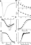
Functional effects of the mutation L1330F. A, Average normalized macropatch current traces elicited with a depolarizing step to −20 mV in Nav1.2–L1330F-transfected neurons (solid line, bipolar and pyramidal neurons pooled); average wild-type Nav1.2 traces are shown for comparison (dashed line, bipolar and pyramidal neurons pooled). Horizontal calibration bar, 500 μs. B, Top, Semilogarithmic plot of the time to half-activation at the indicated potentials for Nav1.2–L1330F (squares) and wild-type Nav1.2 (circles); bottom, time constant of inactivation derived from fits of exponential functions to the decay of the current traces during depolarizations to the indicated potentials. C, Mean current–voltage plots. D, Mean voltage dependence of activation; the lines are mean Boltzmann relationships (solid for L1330F; dashed for wild-type Nav1.2) (Table 1). E, Mean voltage dependence of inactivation; the lines are mean Boltzmann relationships (Table 1). F, Mean kinetics of recovery from inactivation; the lines are mean fits of double-exponential functions to the data (Table 1).
Figure 5.
Figure 5.
Functional effects of the mutation L1563V. A, Average normalized macropatch current traces elicited with a depolarizing step to −20 mV in Nav1.2–L1563V (solid line) and wild-type Nav1.2 (dashed line) transfected neurons. Horizontal calibration bar, 500 μs. B, Top, Semilogarithmic plot of the time to half-activation at the indicated potentials for Nav1.2–L1563V (diamonds) and wild-type Nav1.2 (circles). Statistically significant differences are indicated. Bottom, Time constant of inactivation derived from fits of exponential functions to the decay of the current traces during depolarizations to the indicated potentials. C, Mean current–voltage plots. D, Mean voltage dependence of activation; the lines are mean Boltzmann relationships (solid for L1563V; dashed for wild-type Nav1.2) (Table 1). E, Mean voltage dependence of inactivation; the lines are mean Boltzmann relationships (Table 1). F, Mean kinetics of recovery from inactivation; the lines are mean fits of double-exponential functions to the data (Table 1). *p ≤ 0.05; **p ≤ 0.01.
Figure 6.
Figure 6.
Functional effects of the mutations R223Q and R1319Q studied with classical voltage steps. A, Average normalized macropatch current traces elicited with a depolarizing step to −20 mV in Nav1.2–R223Q (solid line), Nav1.2–R1319Q (dashed-dotted line), and wild-type Nav1.2 (dashed line) transfected neurons. Horizontal calibration bar, 500 μs. B, Top, Semilogarithmic plot of the time to half-activation at the indicated potentials for Nav1.2–R223Q (triangles), Nav1.2–R1319Q (spheres), and wild-type Nav1.2 (circles); the statistical significance is indicated above the points for R1319Q and below the points for R223Q. Bottom, Time constant of inactivation derived from fits of exponential functions to the decay of the current traces during depolarizations to the indicated potentials. C, Mean current–voltage plots. D, Mean voltage dependence of activation; the lines are mean Boltzmann relationships (solid for R223Q, dashed-dotted for R1319Q, and dashed for wild-type Nav1.2) (Table 1). E, Mean voltage dependence of inactivation; the lines are mean Boltzmann relationships (Table 1). F, Mean kinetics of recovery from inactivation; the lines are mean fits of double-exponential functions to the data (Table 1). *p ≤ 0.05; **p ≤ 0.01.
Figure 7.
Figure 7.
Functional effects of the mutations R223Q and R1319Q studied with physiological voltage stimuli. A, Action potential and subthreshold response recorded with sharp microelectrodes in a layer V neuron in neocortical slices. The bottom is the injected depolarizing current pulse. Horizontal calibration bar, 10 ms. B, Currents elicited in transfected neurons by the subthreshold response shown in A. The top shows the subthreshold response used as voltage stimulus; the middle panel shows the recorded subthreshold currents (solid line for R223Q, dashed-dotted line for R1319Q, and dashed line for wild-type Nav1.2). Horizontal calibration bar, 1 ms. The bar graph in the bottom shows the comparison between the area subtended by the subthreshold currents (Table 2). C, Currents elicited in transfected neurons by the action potential shown in A. The top shows the action potential used as voltage stimulus; the middle shows the recorded action currents (solid line for R223Q, dashed-dotted line for R1319Q, and dashed line for wild-type Nav1.2). Horizontal calibration bar, 1 ms. The bar graph in the bottom shows the comparison between the area subtended by the action currents recorded in the three conditions (Table 2). wt, Wild type. *p < 0.05; **p < 0.01.

References

    1. Avanzini G, Franceschetti S. Cellular biology of epileptogenesis. Lancet Neurol. 2003;2:33–42. - PubMed
    1. Baroudi G, Carbonneau E, Pouliot V, Chahine M. SCN5A mutation (T1620M) causing Brugada syndrome exhibits different phenotypes when expressed in Xenopus oocytes and mammalian cells. FEBS Lett. 2000;467:12–16. - PubMed
    1. Berkovic SF, Heron SE, Giordano L, Marini C, Guerrini R, Kaplan RE, Gambardella A, Steinlein OK, Grinton BE, Dean JT, Bordo L, Hodgson BL, Yamamoto T, Mulley JC, Zara F, Scheffer IE. Benign familial neonatal-infantile seizures: characterization of a new sodium channelopathy. Ann Neurol. 2004;55:550–557. - PubMed
    1. Bezanilla F. The voltage sensor in voltage-dependent ion channels. Physiol Rev. 2000;80:555–592. - PubMed
    1. Catterall WA. From ionic currents to molecular mechanisms: the structure and function of voltage-gated sodium channels. Neuron. 2000;26:13–25. - PubMed

Publication types

LinkOut - more resources