Comparative Study
. 2006 Oct 4;26(40):10199-208.
doi: 10.1523/JNEUROSCI.1223-06.2006.

Age-independent synaptogenesis by phosphoinositide 3 kinase

Affiliations
Comparative Study

Age-independent synaptogenesis by phosphoinositide 3 kinase

Alfonso Martín-Peña et al. J Neurosci. .

Abstract

Synapses are specialized communication points between neurons, and their number is a major determinant of cognitive abilities. These dynamic structures undergo developmental- and activity-dependent changes. During brain aging and certain diseases, synapses are gradually lost, causing mental decline. It is, thus, critical to identify the molecular mechanisms controlling synapse number. We show here that the levels of phosphoinositide 3 kinase (PI3K) regulate synapse number in both Drosophila larval motor neurons and adult brain projection neurons. The supernumerary synapses induced by PI3K overexpression are functional and elicit changes in behavior. Remarkably, PI3K activation induces synaptogenesis in aged adult neurons as well. We demonstrate that persistent PI3K activity is necessary for synapse maintenance. We also report that PI3K controls the expression and localization of synaptic markers in human neuroblastoma cells, suggesting that PI3K synaptogenic activity is conserved in humans. Thus, we propose that PI3K stimulation can be applied to prevent or delay synapse loss in normal aging and in neurological disorders.


Figures

Figure 1.
Figure 1.
PI3K regulates synapse number in larval motor neurons. a–g, Larval neuromuscular junctions of muscle fibers F12 or F13 stained with anti-HRP (red) and the synapse-specific nc82 (green) antibodies in control flies (a; Gal4–D42/+) or flies carrying constructs that activate PI3K (b, g; Gal4–D42/UAS-PI3K92E), activate Akt (c; Gal4–D42/UAS–Akt), repress PI3K (d; Gal4–D42/UAS–PI3KDN), and activate GSK3 (f; Gal4–D42/UAS–GSK3) or are homozygous mutants for Akt (e; Akt104226). h, Absolute and relative number of synapses in control flies (D42) or in flies with altered PI3K signaling activity. Numbers inside histograms indicate the sample number per genotype. Asterisks indicate statistically significant differences for all figures. Arrow in g indicates a synaptic bouton with several nc82-positive synapses. Scale bar: a–f, 20 μm; g, 8 μm.
Figure 2.
Figure 2.
PI3K-induced neuromuscular junction synapses are functional. a, Number of synapses, as revealed by nc82 antibody, in muscle fibers 6 and 7. b, Mean frequency of spontaneous mepps at muscle fibers F6 and F7 from late third-instar larvae, indicative of spontaneous neurotransmitter release, is 40% higher for upregulated PI3K larval fibers (Gal4–D42/UAS–PI3K92E) than for control fibers (Gal4–D42) (p = 0.01; n = 12). c, Mean amplitude of the mepps for upregulated PI3K fibers is 40% higher than for controls (p < 0.01; n = 12). d, Recordings from normal (+) and PI3K overexpressing neuromuscular junctions 6–7. e, Frequency distribution of mepps for upregulated PI3K fibers is shifted to higher frequencies and broader. f, The amplitude distribution of the mepps for upregulated PI3K fibers has the same median as the controls but is broader and with a more important tail at high amplitudes. g, Evoked EPSPs for upregulated PI3K fibers show a significant 9.4 mV increase in amplitude (p < 0.001; n = 14) but the same rise and tail as controls.
Figure 3.
Figure 3.
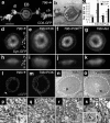
PI3K regulates synapse number in adult central neurons. a, Frontal view of a whole-mount adult brain showing 36 neurons [cell bodies (cb)] projecting to the EB from each side and receiving their input through a dendritic tuft (dt) (Gal4–796/+; UAS–CD8–GFP/+). b, Plastic-embedded section of the EB visualized with anti-β-Gal antibody (Gal4–796/+; UAS–LacZ/+) showing the glomerular organization of the EB neuropil (arrows). c, Effects of PI3K and Akt activity on the relative synaptic volume (syn-GFP) and cell size (cyto-GFP) of EB neurons. Values are normalized with respect to controls (Gal4–796/+; GFP/+). d–g, Confocal serial reconstructions of EBs to measure synaptic volume with syn-GFP (Gal4–796/UAS–syn-GFP) from control (+), activated PI3K (UAS–PI3K), repressed PI3K (UAS–PI3KDN), and activated Akt (UAS–Akt) flies. PI3K and Akt increase EB size, whereas PI3KDN slightly reduces it. h–k, Top view of EBs corresponding to previous genotypes. l, m, Single R4m neurons expressing CD8–GFP (Gal4–796/UAS–CD8–GFP) visualized with anti-GFP from control (+) and UAS–PI3K brains. Cell size changes are noticed in the diameter of axons (l, arrow) and the crown-shaped projection. n, o, Low-magnification TEM sections showing EB (dotted circles) from control (same as d) and activated PI3K (same as e) brains. p, q, High magnification of insets shown in n and o. The arrowheads indicate typical synapse specialization (inset, arrowhead). r, s, Dendritic tuft from Gal4–796/+; UAS–PI3K/UAS–HRP neurons. HRP deposits label the cell profiles in dark (r, arrowheads). A higher magnification is shown in s. Inset in s shows reciprocal synapses between HRP-positive (black arrowhead) and HRP-negative (white arrowhead) neuronal profiles. Scale bar: a, b, 40 μm; d–k, 20 μm. A, Anterior; D, dorsal; P, posterior; V, ventral.
Figure 4.
Figure 4.
Characterization of the PI3K synaptogenic effect. a, The developmental profile of EB synaptic volume (Gal4–796/UAS–syn-GFP) is similar in control (+) and PI3K-overexpressing (UAS–PI3K) flies. The EB size difference is detected at all ages. b–d, The locomotion assay in the Buridan’s arena shows increased active time (b), walked distance (c), and the number of full walks between landmarks in adult flies expressing PI3K compared with controls (genotypes as in a). The expression of PI3KDN yields opposite effects. Movement speed (e) remains normal in PI3K but reduced in PI3KDN animals.
Figure 5.
Figure 5.
Synaptogenic effect of other signaling components. a–c, Relative synaptic (Gal4–796/UAS–syn-GFP) and cellular (Gal4–796/UAS–GFP) volume of EB overexpressing components of the PI3K signaling pathway. a, Rheb overexpression increases the synaptic domain, whereas GSK3 significantly decreases it. b, Changes in InR (InR and InRDN overexpression) and Chico activity affect synapse number (but see Results). c, EgfR activity (EgfR and EgfRDN overexpression) affects cell size but not synapse number. The number of brains analyzed per genotype is indicated on each histogram.
Figure 6.
Figure 6.
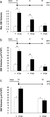
PI3K activity is necessary for synapse maintenance. a, b, Relative (a) and absolute (b) synaptic volume of the EB (Gal4–796/UAS–syn-GFP; Gal80ts/+) in control (+) or PI3K-expressing (UAS–PI3K) flies in which Gal4 activity was switched off in 8-d-old adult flies. Flies were raised at 30°C until adults reached 8 d. This temperature inhibits Gal80ts, allowing Gal4 activity. Then, the culture temperature was shifted to 17°C, allowing Gal80ts to repress Gal4 activity (switch-off). Syn-GFP was measured on days 8, 9, and 10. c, Equivalent experiments switching off the Gal4–796 expression at day 15 and measuring at day 18. The synaptic volume decreases dramatically in PI3K flies, indicating that the ectopic synapses require constant PI3K stimulation.
Figure 7.
Figure 7.
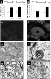
PI3K is sufficient for synapse formation and it protects aging neurons. a, Relative synaptic volume of the EB (Gal4–796/UAS–syn-GFP; Gal80ts/+) in control (+) or PI3K-expressing (UAS–PI3K) flies in which Gal4 activity was switched on in 15- or 30-d-old adult flies. Flies were raised at 17°C until adults reached 15 or 30 d (top). This temperature allows Gal80ts to repress Gal4 activity (no expression). Then, the culture temperature was shifted to 30°C, inhibiting Gal80ts and allowing Gal4 activity (switch-on). syn-GFP was measured at least 15 d later. Flies that start expressing PI3K at days 15 (left) or 30 (right) induce significant number of neo-synapses when analyzed at days 40 and 45, respectively. b, Absolute volume of the EB from brains subjected to switch-on experiments but monitoring synapses by the nc82 antibody. c, d, Frontal views of EBs seen as stacks of confocal planes from an experiment as in b visualized at day 35. Note the higher synaptic signal in the PI3K brain (d) with respect to control (c). e, Toluidine blue-stained semithin section of the EB of a 50-d-old adult subjected to PI3K switch-on at day 30. Age-related vacuoles are abundant in areas adjacent to the EB (arrowheads) and in a crown within the EB (black arrow). f, Anomalous electron-dense bodies characteristic of aged neurons. g, High-magnification TEM corresponding to the square in e showing age-characteristic disorganization of the cytoskeleton (stars) and the general depletion of electron-dense materials. h, High-magnification TEM of the circle in e showing abundance of small synaptic vesicles and multiple synapses converging on a single dendrite (arrowheads). Age-related electron-dense bodies (arrow) can also be found in some neurons.
Figure 8.
Figure 8.
PI3K regulates expression and localization of synaptic markers in human neuroblastoma cells. a, Distribution of the synaptic vesicle marker SV2 (green) in normal SH-SY5Y cultured cells. b, SH-SY5Y cells incubated with 50 μg/ml 740Y-P induce the translocation of SV2 to the projections. c, d, 740Y-P induces higher expression levels of SV2 and higher levels of phospho-Akt as detected in Western blot. LY294002 (5 μm) and wortmannin (10 μm) reduce SV2 levels by 70% and phosphorylated Akt by 30%. Phospho-Akt levels are measured with respect to total Akt, and SV2 levels are normalized versus β-tubulin. White histograms, Control culture; black histograms, treated culture.

Similar articles

Cited by

References

    1. Abbott MA, Wells DG, Fallon JR. The insulin receptor tyrosine kinase substrate p58/53 and the insulin receptor are components of CNS synapses. J Neurosci. 1999;19:7300–7308. - PMC - PubMed
    1. Acebes A, Ferrus A. Increasing the number of synapses modifies olfactory perception in Drosophila. J Neurosci. 2001;21:6264–6273. - PMC - PubMed
    1. Agarwal-Mawal A, Qureshi HY, Cafferty PW, Yuan Z, Han D, Lin R, Paudel HK. 14–3-3 connects glycogen synthase kinase-3 beta to tau within a brain microtubule-associated tau phosphorylation complex. J Biol Chem. 2003;278:12722–12728. - PubMed
    1. Arendt T, Gartner U, Seeger G, Barmashenko G, Palm K, Mittmann T, Yan L, Hummeke M, Behrbohm J, Bruckner MK, Holzer M, Wahle P, Heumann R. Neuronal activation of Ras regulates synaptic connectivity. Eur J Neurosci. 2004;19:2953–2966. - PubMed
    1. Barth M, Hirsch HV, Meinertzhagen IA, Heisenberg M. Experience-dependent developmental plasticity in the optic lobe of Drosophila melanogaster. J Neurosci. 1997;17:1493–1504. - PMC - PubMed

Publication types

Substances