

The emergence of radioimmunoscintigraphy for prostate cancer
- PMID: 17021623
- PMCID: PMC1578531
The emergence of radioimmunoscintigraphy for prostate cancer
Abstract
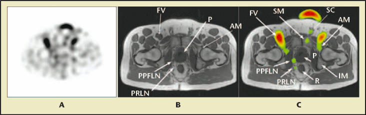
The ability to label tissue-specific antibodies has long been of interest for improving detection and guidance for therapeutic applications. The most studied target for prostate cancer is the prostate-specific membrane antigen, which is upregulated in prostate cancer, hormone-refractive disease, and prostate cancer metastases. Investigations using radioimmunoscintigraphy with the radiolabeled 7E11 antibody capromab pendetide have significantly improved sensitivity for prostate cancer detection compared with standard cross-sectional imaging, based on tissue confirmation of pathologic results. Over the past 5 years, significantly greater image resolution from improved camera technology and the use of co-registration to fuse functional and anatomic (computerized tomography and magnetic resonance imaging) images have dramatically enhanced prostate cancer localization. Outcomes data from several sources have spurred a resurgence in interest in this imaging modality.
Figures


Similar articles
-
Update on fused capromab pendetide imaging of prostate cancer.Clin Prostate Cancer. 2005 Mar;3(4):230-8. doi: 10.3816/cgc.2005.n.004. Clin Prostate Cancer. 2005. PMID: 15882479 Review.
-
Clinical Applications of Radioimmunoscintigraphy With Prostate-Specific Antibodies for Prostate Cancer.Cancer Control. 1998 Nov;5(6):493-499. doi: 10.1177/107327489800500601. Cancer Control. 1998. PMID: 10761097
-
Utility of capromab pendetide (ProstaScint) imaging in the management of prostate cancer.Tech Urol. 2001 Mar;7(1):27-37. Tech Urol. 2001. PMID: 11272670 Review.
-
Capromab pendetide. A review of its use as an imaging agent in prostate cancer.Drugs Aging. 1998 Apr;12(4):293-304. doi: 10.2165/00002512-199812040-00004. Drugs Aging. 1998. PMID: 9571393 Review.
-
Prostate cancer abdominal metastases detected with indium-111 capromab pendetide.J Nucl Med. 1998 Apr;39(4):650-2. J Nucl Med. 1998. PMID: 9544673
Cited by
-
Antibody-based imaging of HER-2: moving into the clinic.Curr Mol Med. 2013 Dec;13(10):1523-37. doi: 10.2174/1566524013666131111120951. Curr Mol Med. 2013. PMID: 24206138 Free PMC article. Review.
-
Recent trends in antibody-based oncologic imaging.Cancer Lett. 2012 Feb 28;315(2):97-111. doi: 10.1016/j.canlet.2011.10.017. Epub 2011 Oct 20. Cancer Lett. 2012. PMID: 22104729 Free PMC article. Review.
-
N-[N-[(S)-1,3-Dicarboxypropyl]carbamoyl]-4-[18F]fluorobenzyl-L-cysteine, [18F]DCFBC: a new imaging probe for prostate cancer.Clin Cancer Res. 2008 May 15;14(10):3036-43. doi: 10.1158/1078-0432.CCR-07-1517. Clin Cancer Res. 2008. PMID: 18483369 Free PMC article.
References
-
- Jemal A, Murray T, Ward E, et al. Cancer statistics, 2005. CA Cancer J Clin. 2005;55:10–30. - PubMed
-
- Khan MA, Partin AW, Mangold LA, et al. Probability of biochemical recurrence by analysis of pathologic stage, Gleason score, and margin status for localized prostate cancer. Urology. 2003;62:866–871. - PubMed
-
- Kattan MW, Eastham JA, Wheeler TM, et al. Counseling men with prostate cancer: a nomogram for predicting the presence of small, moderately differentiated, confined tumors. J Urol. 2003;170:1792–1797. - PubMed
-
- Murray SK, Breau RH, Guha AK, Gupta R. Spread of prostate carcinoma to the perirectal lymph node basin: analysis of 112 rectal resections over a 10-year span for primary rectal adenocarcinoma. Am J Surg Pathol. 2004;28:1154–1162. - PubMed
-
- Saitoh H, Yoshida K-I, Uchijima Y, et al. Two different lymph node metastatic patterns of a prostatic cancer. Cancer. 1990;65:1843–1846. - PubMed
LinkOut - more resources
Full Text Sources
Miscellaneous