

Evidence supporting extraocular muscle pulleys: refuting the platygean view of extraocular muscle mechanics
- PMID: 17022164
- PMCID: PMC1858665
- DOI: 10.3928/01913913-20060901-05
Evidence supporting extraocular muscle pulleys: refuting the platygean view of extraocular muscle mechanics
Abstract
Background: Late in the 20th century, it was recognized that connective tissue structures in the orbit influence the paths of the extraocular muscles and constitute their functional origins. Targeted investigations of these connective tissue "pulleys" led to the formulation of the active pulley hypothesis, which proposes that pulling directions of the rectus extraocular muscles are actively controlled via connective tissues.
Purpose: This review rebuts a series of criticisms of the active pulley hypothesis published by Jampel, and Jampel and Shi, in which these authors have disputed the existence and function of the pulleys.
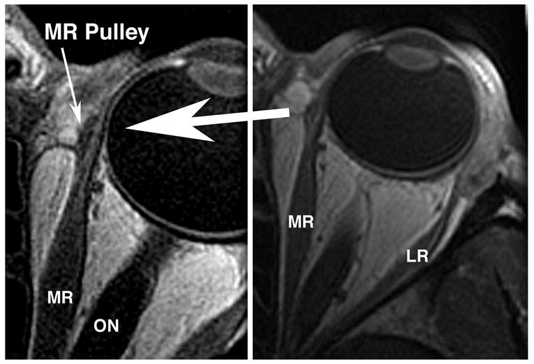
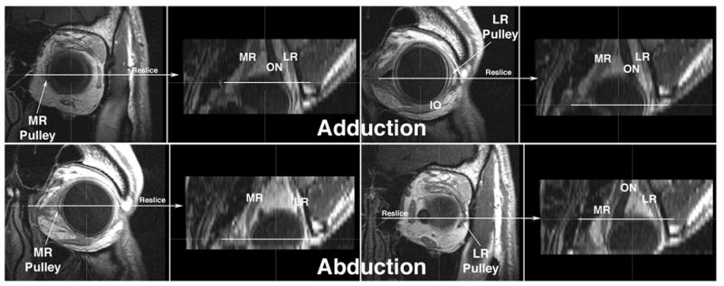
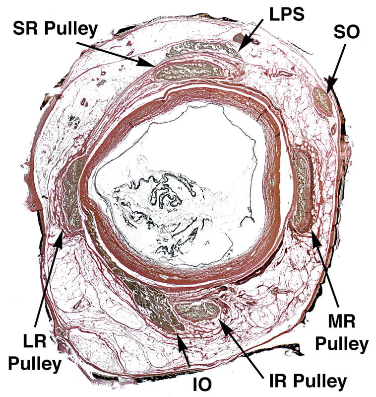
Methods: This article reviews published evidence for the existence of orbital pulleys, the active pulley hypothesis, and physiological tests of the active pulley hypothesis. Magnetic resonance imaging in a living subject and histological examination of a human cadaver directly illustrate the relationship of pulleys to extraocular muscles.
Results: Strong scientific evidence is cited that supports the existence of orbital pulleys and their role in ocular motility. The criticisms of the hypothesis have ignored mathematical truisms and strong scientific evidence.
Conclusions: Actively control led orbital pulleys play a fundamental role in ocular motility. Pulleys profoundly influence the neural commands required to control eye movements and binocular alignment. Familiarity with the anatomy and physiology of the pulleys is requisite for a rational approach to diagnosing and treating strabismus using emerging methods. Conversely, approaches that deny or ignore the pulleys risk the sorts of errors that arise in geography and navigation from incorrect assumptions such as those of a flat ("platygean") earth.
Figures
Comment in
-
The active pulley hypothesis.J Pediatr Ophthalmol Strabismus. 2006 Sep-Oct;43(5):272. doi: 10.3928/01913913-20060901-12. J Pediatr Ophthalmol Strabismus. 2006. PMID: 17022159 No abstract available.
Similar articles
-
Functional anatomy of the orbit in strabismus surgery: Connective tissues, pulleys, and the modern surgical implications of the "arc of contact" paradigm.J Anat. 2024 Jun;244(6):887-899. doi: 10.1111/joa.14009. Epub 2024 Jan 19. J Anat. 2024. PMID: 38243145 Free PMC article. Review.
-
Location and stability of rectus muscle pulleys. Muscle paths as a function of gaze.Invest Ophthalmol Vis Sci. 1997 Jan;38(1):227-40. Invest Ophthalmol Vis Sci. 1997. PMID: 9008649
-
Evidence for active control of rectus extraocular muscle pulleys.Invest Ophthalmol Vis Sci. 2000 May;41(6):1280-90. Invest Ophthalmol Vis Sci. 2000. PMID: 10798641
-
Evidence against mobile pulleys on the rectus muscles and inferior oblique muscle: central nervous system controls ocular kinematics.J Pediatr Ophthalmol Strabismus. 2006 Sep-Oct;43(5):289-95. doi: 10.3928/01913913-20060901-04. J Pediatr Ophthalmol Strabismus. 2006. PMID: 17022163 Review.
-
Evidence for fibromuscular pulleys of the recti extraocular muscles.Invest Ophthalmol Vis Sci. 1995 May;36(6):1125-36. Invest Ophthalmol Vis Sci. 1995. PMID: 7730022
Cited by
-
Minimally invasive surgery for thyroid eye disease.Indian J Ophthalmol. 2015 Nov;63(11):847-53. doi: 10.4103/0301-4738.171967. Indian J Ophthalmol. 2015. PMID: 26669337 Free PMC article.
-
Postnatal changes in the developing rat extraocular muscles.Invest Ophthalmol Vis Sci. 2011 Jun 7;52(7):3962-9. doi: 10.1167/iovs.10-6866. Invest Ophthalmol Vis Sci. 2011. PMID: 21372011 Free PMC article.
-
Extraocular Muscles Tension, Tonus, and Proprioception in Infantile Strabismus: Role of the Oculomotor System in the Pathogenesis of Infantile Strabismus-Review of the Literature.Scientifica (Cairo). 2016;2016:5790981. doi: 10.1155/2016/5790981. Epub 2016 Feb 23. Scientifica (Cairo). 2016. PMID: 27006860 Free PMC article. Review.
-
Fascicular specialization in human and monkey rectus muscles: evidence for anatomic independence of global and orbital layers.Invest Ophthalmol Vis Sci. 2007 Jul;48(7):3089-97. doi: 10.1167/iovs.06-0692. Invest Ophthalmol Vis Sci. 2007. PMID: 17591878 Free PMC article.
-
Dynamics of primate oculomotor plant revealed by effects of abducens microstimulation.J Neurophysiol. 2009 Jun;101(6):2907-23. doi: 10.1152/jn.91045.2008. Epub 2009 Mar 18. J Neurophysiol. 2009. PMID: 19297512 Free PMC article.
References
-
- Jampel RS, Shi DX. Evidence against mobile pulleys on the rectus muscles and inferior oblique muscle: Central nervous system controls ocular kinematics. J Peadiatr Ophthalmol Strabismus. 2006;43 in press. - PubMed
-
- Jampel RS. Pulley and globe stability. Invest Ophthalmol Vis Sci. 2006 e-letter.
-
- Jampel RS. The superior rectus is not coupled to the superior oblique pulley. Invest Ophthalmol Vis Sci e-letter. 2006
-
- Demer JL. Author response: The superior rectus is not coupled to the superior oblique pulley. Invest Ophthalmol Vis Sci. 2006 e-letter.
Publication types
MeSH terms
Grants and funding
LinkOut - more resources
Full Text Sources
