Review
. 2006 Oct;5(10):2505-22.
doi: 10.1021/pr0603349.

Nanotools for megaproblems: probing protein misfolding diseases using nanomedicine modus operandi

Affiliations
Review

Nanotools for megaproblems: probing protein misfolding diseases using nanomedicine modus operandi

Vladimir N Uversky et al. J Proteome Res. 2006 Oct.

Abstract

Misfolding and self-assembly of proteins in nanoaggregates of different sizes and morphologies (nanoensembles, primary nanofilaments, nanorings, filaments, protofibrils, fibrils, etc.) is a common theme unifying a number of human pathologies termed protein misfolding diseases. Recent studies highlight increasing recognition of the public health importance of protein misfolding diseases, including various neurodegenerative disorders and amyloidoses. It is understood now that the first essential elements in the vast majority of neurodegenerative processes are misfolded and aggregated proteins. Altogether, the accumulation of abnormal protein nanoensembles exerts toxicity by disrupting intracellular transport, overwhelming protein degradation pathways, and/or disturbing vital cell functions. In addition, the formation of inclusion bodies is known to represent a major problem in the production of recombinant therapeutic proteins. Formulation of these therapeutic proteins into delivery systems and their in vivo delivery are often complicated by protein association. Thus, protein folding abnormalities and subsequent events underlie a multitude of human pathologies and difficulties with protein therapeutic applications. The field of medicine therefore can be greatly advanced by establishing a fundamental understanding of key factors leading to misfolding and self-assembly responsible for various protein folding pathologies. This article overviews protein misfolding diseases and outlines some novel and advanced nanotechnologies, including nanoimaging techniques, nanotoolboxes and nanocontainers, complemented by appropriate ensemble techniques, all focused on the ultimate goal to establish etiology and to diagnose, prevent, and cure these devastating disorders.


Figures

Figure 1
Figure 1
Modern, hierarchical view of protein misfolding and amyloid fibrils: intra- and extracellular inclusions in different tissues associated with misfolding diseases (A) are proteinaceous deposits (B). These deposits are comprised of amyloid fibrils, crude structures which can be resolved by EM or AFM (C). Cryo-EM analysis provides a detailed view of fibrils as an intertwined bundle of several filaments (D). Modeling based on X-ray diffraction and EM/AFM analyses gives the ultrastructure of amyloid fibrils and filaments as cross-β spines (E). The first structure of a model amyloid fibril comprised of a short peptide derived from the yeast prion protein was determined at atomic resolution (F) (for more details see Figure 3). There are at least 20 different proteins associated with protein misfolding diseases. An example of one of the amyloidogenic polypeptides, bovine prion protein, is shown in panel G. Panels D and E are modified from ref with the permission of authors. Panel F is adapted with permission from ref . Copyright 2005 Macmillan Publishers Ltd: Nature. Panel G is a generous gift of Prof. Wutrich.
Figure 2
Figure 2
Individual protein molecules can be physically separated via encapsulation in silica glass using the sol–gel technique.
Figure 3
Figure 3
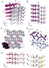
Structure of a GNNQQNY-based amyloid fibril. (A) The pair-of-sheets structure, showing the backbone of each β-strand as an arrow, with side chains protruding. The dry interface is between the two sheets, with the wet interfaces on the outside surfaces. The side chains of Asn2, Gln4, and Asn6 point inward, forming the dry interface. The 21 screw axis of the crystal is shown as the vertical line. It rotates one of the strands of the near sheet 180°about the axis and moves it up 4.87 Å so that it is superimposed on one of the strands of the far sheet. (B) The steric zipper viewed edge on (down the a axis). Note the vertical shift of one sheet relative to the other, allowing interdigitation of the side chains emanating from each sheet. The amide stacks of the dry interface are shaded in gray at the center, and those of the wet interface are shaded in pale red on either side. (C) The GNNQQNY crystal viewed down the sheets (from the top of panel A, along the b axis). Six rows of β-sheets run horizontally. Peptide molecules are shown in black, and water molecules are red plus signs. The atoms in the lower left unit cell are shown as spheres representing van der Waals radii. (D) The steric zipper. This is a close-up view of a pair of GNNQQNY molecules from the same view as panel C, showing the remarkable shape complementarity of the Asn and Gln side chains protruding into the dry interface. 2Fo – Fc electron density is shown, and the position of the central screw axis is indicated. (E) Views of the β-sheets from the side (down the c axis), showing three β-strands with the inter-strand hydrogen bonds. Side-chain carbon atoms are yellow. Backbone hydrogen bonds are shown by purple or gray dots and side-chain hydrogen bonds by yellow dots. Hydrogen bond lengths are noted in angstroms. The views of the interfaces are close to the views of panel A. The left-hand set is viewed from the center of the dry interface; the right-hand set is viewed from the wet interface. Note the amide stacks in both interfaces. Reprinted with permission from ref . Copyright 2005 Macmillan Publishers Ltd: Nature.
Figure 4
Figure 4
Nanocontainers for protein delivery. The core represents the polyion complex of proteins and synthetic polyelectrolytes of opposite charge. The PEO–PPO–PEO chains grafted to the polyelectrolyte within the core form a micelle-like structure around the core, with the hydrophobic PPO blocks near the core and the hydrophilic PEO blocks forming the exterior shell. Additional nonmodified PEO–PPO–PEO chains fill in to optimize the stability of the micelle-like structure.
Figure 5
Figure 5
Intracellular localization of native HRP (A) and HRP-P85 (B) in living bovine brain microvessel endothelial cells (BBMEC) visualized by confocal laser scanning microscopy. HRP was labeled with Alexa Fluor 594.

References

    1. Cohen AS. General introduction and a brief history of the amyloid fibril. In: Marrink J, Van Rijswijk MH, editors. Amyloidosis. Martinus Nijhoff; Dordrecht, The Netherlands: 1986. pp. 3–19.
    1. Virchow R. Cellulose-Frage. Virchows Arch Pathol Anat. 1854;6:416–426.
    1. Friedreich NK, Zur Amyloidfrage A. Virchows Arch Pathol Anat. 1859;15:50–65.
    1. Hanssen O. Ein Beitrag zur Chemie der amyloiden Entartung. Biochem Z. 1908;13:185–198.
    1. Alzheimer A. Über eine eigenartige Eskrankung der Nirnrinde. Allg Z Psychiatr Psych-Gerichtl. 1907;64:146–148.

Publication types

MeSH terms