. 2007 Jan;148(1):116-27.
doi: 10.1210/en.2006-0561. Epub 2006 Oct 5.

Exposure to environmentally relevant doses of the xenoestrogen bisphenol-A alters development of the fetal mouse mammary gland

Affiliations

Exposure to environmentally relevant doses of the xenoestrogen bisphenol-A alters development of the fetal mouse mammary gland

Laura N Vandenberg et al. Endocrinology. 2007 Jan.

Abstract

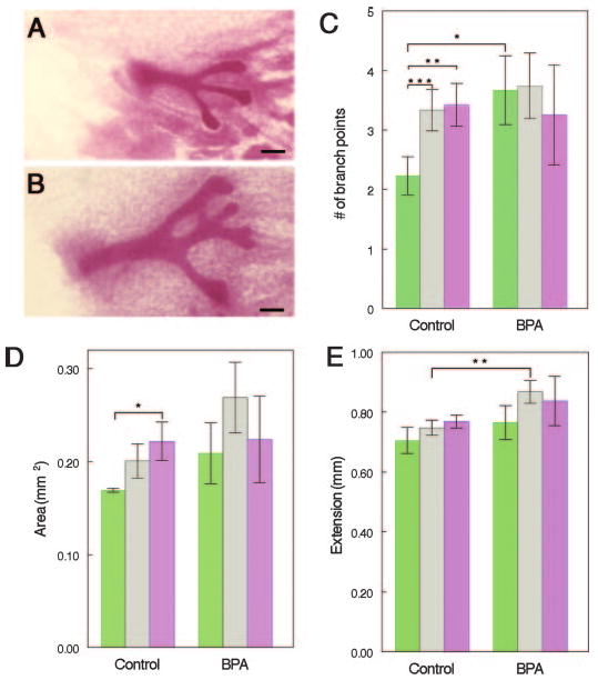
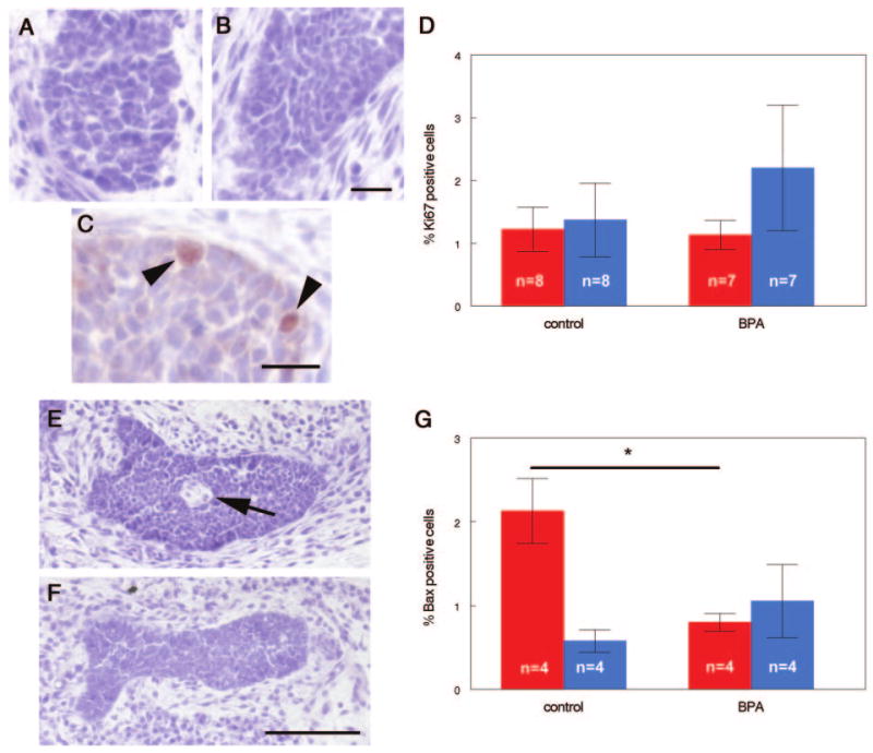
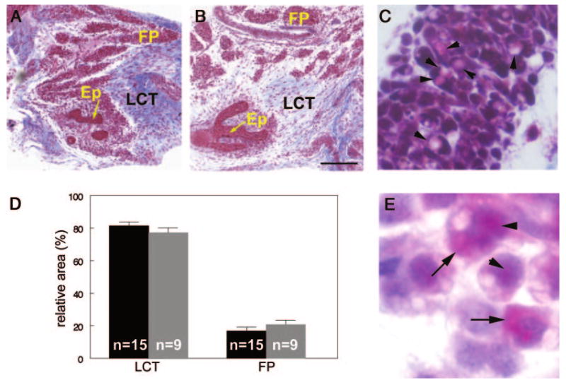
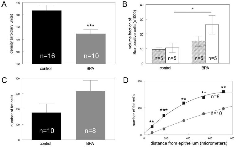
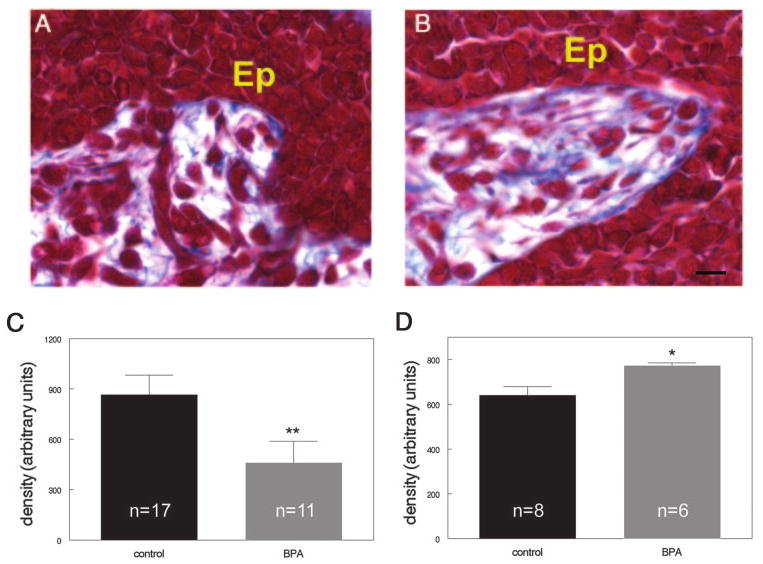
Humans are routinely exposed to bisphenol-A (BPA), an estrogenic compound that leaches from dental materials, food and beverage containers, and other plastic consumer products. Effects of perinatal BPA exposure on the mouse mammary gland have been observed in puberty and adulthood, long after the period of exposure has ended. The aim of this study was to examine fetal mammary gland development at embryonic day (E)18 and assess changes in the tissue organization and histoarchitecture after exposure to an environmentally relevant dose of BPA. In unexposed fetuses, the relative position of the fetus with respect to its female and male siblings in the uterus influenced growth of the ductal tree, which was more developed in females placed between two males than in females placed between two females. Exposure of dams to 250 ng BPA per kilogram body weight per day from E8 to E18 significantly increased ductal area and ductal extension in exposed fetuses and obliterated positional differences. In the stroma, BPA exposure promoted maturation of the fat pad and altered the localization of collagen. Within the epithelium, BPA exposure led to a decrease in cell size and delayed lumen formation. Because mammary gland development is dependent on reciprocal interactions between these compartments, the advanced maturation of the fat pad and changes in the extracellular matrix may be responsible for the altered growth, cell size, and lumen formation observed in the epithelium. These results suggest that alterations in mammary gland phenotypes observed at puberty and adulthood in perinatally exposed mice have their origins in fetal development.


Figures

Fig. 1
Fig. 1
Effects of intrauterine position on the growth of the mammary ductal tree in control and BPA-exposed fetuses. A, Whole mount of control 0M female mammary gland. B, Whole mount of BPA-exposed 0M female mammary gland. Note the increased size of the epithelial cords, compared with controls. C, Number of branching points was increased in control 1M (gray) and 2M (violet) females, compared with 0M (green) females. In BPA-exposed females, 0M females were as developed as 1M and 2M females. D, Area subtended by the ductal tree was increased in control 2M females, compared with 0M females. These position-specific effects disappeared after BPA exposure. E, Ductal extension was significantly increased in 1M females exposed to BPA, compared with 1M control females. Scale bar, 100 μm. n = 8–15/positional group for both treatments. *, P < 0.045, **, P < 0.03, ***, P < 0.025.
Fig. 2
Fig. 2
Development of the mammary epithelium in control and BPA-exposed females. Epithelial cells are larger and more heterogeneous in shape and size in control animals (A), compared with BPA-exposed (B). C, Ki67 expression in epithelium from a control fetus (arrowheads indicate positive cells). D, Distribution of Ki67-positive cells in the inner (red) and outer cord cells (blue) was similar for both treatments. E, Lumen formation (arrow) was observed in 38% of control animals but in none of the BPA-exposed animals (F). G, Expression of proapoptotic marker Bax was significantly decreased in the inner cord cells (red) of BPA-exposed relative to controls, but there were no differences in Bax expression in the outer cord cells (blue) by treatment. Scale bars (B and C), 20 μm; (F), 100 μm. *, P = 0.021.
Fig. 3
Fig. 3
Appearance of the mammary stroma in control and BPA-exposed females. Masson's Trichrome staining of control (A) and BPA-exposed (B) E18 mammary glands. C, Clear vacuoles (arrowheads) are observed in some cells within the developing fat pad. D, The relative area of loose connective tissue and fat pad are similar in control (black) and BPA-exposed (gray) mammary glands. E, Localization of ALBP in cells of the fat pad in the nuclear (arrowheads) and cytoplasmic (arrows) compartments of adipocytes. Ep, Epithelium, FP, fat pad, LCT, loose connective tissue. Scale bar, 200 μm.
Fig. 4
Fig. 4
Alterations are observed in the developing fat pad of BPA-exposed females, compared with controls. A, The OD of the fat pad in BPA-exposed animals (gray) is decreased, compared with controls (black). B, Expression of the proapoptotic marker Bax is significantly increased in the developing fat pad (white) in BPA-exposed animals relative to controls. No statistically significant differences were noted in the loose connective tissue (shaded). C, The total number of cells containing fat vacuoles was increased in BPA-exposed females, compared with controls, although this increase was not significant. D, The number of adipocytes was significantly increased within 1 mm from the developing epithelium in BPA-exposed females (squares), compared with controls (circles). *, P < 0.05; **, P < 0.02; ***, P < 0.005.
Fig. 5
Fig. 5
BPA exposure alters collagen localization in the fetal mammary gland. Masson's Trichrome staining of control (A) and BPA-exposed (B) E18 mammary glands. Note the increase in blue collagen fibers directly abutting the epithelium in BPA-exposed mammary glands. C, The density of collagen in the entire stromal compartment is significantly decreased in BPA-exposed females, compared with controls. D, The density of collagen within 10 μm of the epithelial ducts is significantly increased in BPA-exposed females. [Ep, Epithelium; blue, collagen fibers. Scale bar, 10 μm]. *, P = 0.042; **, P = 0.010.
Fig. 6
Fig. 6
ERα is expressed in the E18 mammary gland. A, ERα expression in the developing fat pad. Inset shows higher magnification (arrows indicate positive cells). B, Punctate expression of ERα (arrows indicate positive cells) is observed in some epithelial cords at E18. Inset shows higher magnification of positive cells. C, Negative control, E18 mammary gland treated in the same way as A and B but without the addition of primary antibody. Scale bar, 100 μm.

References

    1. Barker DJP. The developmental origins of adult disease. Eur J Endocrinol. 2003;18:733–736. - PubMed
    1. Godfrey KM, Barker DJP. Fetal programming and adult health. Public Health Nutr. 2001;4:611–624. - PubMed
    1. Markey CM, Luque EH, Munoz de Toro MM, Sonnenschein C, Soto AM. In utero exposure to bisphenol A alters the development and tissue organization of the mouse mammary gland. Biol Reprod. 2001;65:1215–1223. - PubMed
    1. Munoz de Toro MM, Markey CM, Wadia PR, Luque EH, Rubin BS, Sonnenschein C, Soto AM. Perinatal exposure to Bisphenol A alters peripubertal mammary gland development in mice. Endocrinology. 2005;146:4138–4147. - PMC - PubMed
    1. Durando M, Kass L, Piva J, Sonnenschein C, Soto AM, Luque EH, Munoz de Toro MM. Prenatal bisphenol A exposure induces preneoplastic lesions in the mammary gland in Wistar rats. Environ Health Perspect. 2006 doi: 10.1289/ehp.9282. - DOI - PMC - PubMed

Publication types

MeSH terms