. 2006 Oct 17;103(42):15663-8.
doi: 10.1073/pnas.0607241103. Epub 2006 Oct 5.

Ionic control of ocular growth and refractive change

Affiliations

Ionic control of ocular growth and refractive change

Sheila G Crewther et al. Proc Natl Acad Sci U S A. .

Abstract

The physiological mechanisms underlying the abnormal vitreal and ocular growth and myopic refractive errors induced under conditions of visual form deprivation in many animal species, including humans, are unknown. This study demonstrates, using energy dispersive x-ray microanalysis, a systematic pattern of changes in the elemental distribution of K, Na, and Cl across the entire retina in experimental form deprivation myopia and in the 5 days required for refractive normalization after occluder removal. In our report we link the ionic environment associated with physiological activity of the retina under a translucent occluder to refractive change and describe large but reversible environmentally driven increases in potassium, sodium, and chloride abundances in the neural retina. Our results are consistent with the notion of ionically driven fluid movements as the vector underlying the myopic increase in ocular size. New treatments for myopia, which currently affects nearly half of the human population, may result.


Conflict of interest statement

The authors declare no conflict of interest.

Figures

Fig. 1.
Fig. 1.
Effect of occlusion on temporal contrast and elemental abundance across the retina. (a) Sample phototransistor output from the two model eyes [one with an occluder (solid line)] under conditions of constant ambient illumination, but with dynamic patterned stimuli provided by chicks moving past the two sensors within the enclosure. The occluder dramatically reduced the temporal contrast. (b) Scanning electron micrograph of retina and choroid of a control eye. The five anatomically defined regions visually selected for assessment were choroid, RPE–SRS–OS, inner segment/outer nuclear layer (IS), inner nuclear layer (INL), and ganglion cell layer (GCL). The white rectangles indicate the irradiated areas (each 37 μm × 26 μm). (Scale bar: 100 μm.) (c) Spectra indicating x-ray counts recorded in the five designated regions from a freeze-dried FD eye (Left) and from the nondeprived fellow eye (Right) at occluder removal (T = 0 h). The major peaks shown from left to right in each figure are Na, P, S, Cl, and K. Note the dramatically raised abundances of Na and Cl in the choroid and outer retinal areas of the FD eye. K abundance was also higher in the outer retina/RPE region of the FD eye. (The scale bar in the bottom right spectrum applies to all spectra and denotes 1 keV horizontally and 50 counts vertically.)
Fig. 2.
Fig. 2.
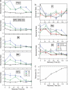
Mean elemental ratios across the retina after form deprivation. (a) Mean elemental ratios (myopic eye:fellow eye) of potassium, sodium, and chlorine at T = 0 and during the first 120 h after occluder removal shown separately for each of the five regions. Regions sampled include choroid (Chor), RPE–SRS–OS, inner segments (IS), inner nuclear layer (INL), and ganglion cell layer (GCL). (b) Elemental ratios (myopic eye:fellow eye) shown separately for K, Na, and Cl over the first 120 h after occluder removal in the inner retina. (c) Time course of refractive state. Refractive recovery of the occluded eyes of chicks is almost linear with time from ≈22 diopters of myopia at occluder removal to zero at ≈5 days of recovery under normal visual stimulus conditions. Error bars represent 1 SE.
Fig. 3.
Fig. 3.
Scatter plots showing the pairwise relationships among Na, K, and Cl abundances (x-ray counts) for the myopic eyes and fellow eyes combining data across all retinal levels and time points. Regression lines (myopic, red line; fellow, blue line) are drawn. (a) A strong correlation exists between Cl and Na [for the myopic eyes (Spearman's ρ = 0.90, P < 0.0001) but not fellow eyes (ρ = 0.04, P > 0.05)]. (b and c) Potassium abundance is held within a smaller range than for Na or Cl, and correlations were <0.4 for both myopic and fellow eyes.

References

    1. Seet B, Wong T, Tan D, Saw S, Balakrishnan , Lee L, Lim A. Br J Ophthalmol. 2001;85:521–526. - PMC - PubMed
    1. Wallman J, Winawer J. Neuron. 2004;43:447–468. - PubMed
    1. Wallman J, Turkel J, Trachtman J. Science. 1978;201:1249–1251. - PubMed
    1. Liang H, Crewther SG, Crewther DP, Junghans BM. Invest Ophthalmol Vis Sci. 2004;45:2463–2474. - PubMed
    1. Liang H, Crewther SG, Crewther DP, Pirie B. Aust NZ J Ophthalmol. 1996;24:41–44. - PubMed

Publication types