. 2007 Mar 1;359(1):233-42.
doi: 10.1016/j.virol.2006.09.009. Epub 2006 Oct 9.

Injection of the sciatic nerve with TMEV: a new model for peripheral nerve demyelination

Affiliations

Injection of the sciatic nerve with TMEV: a new model for peripheral nerve demyelination

Kristen M Drescher et al. Virology. .

Abstract

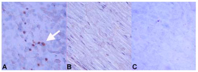
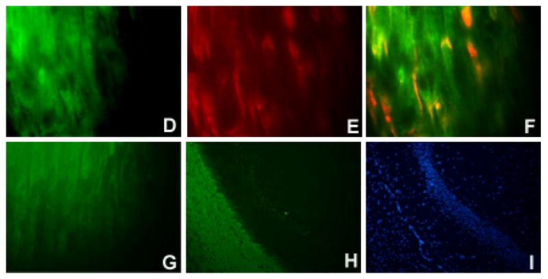
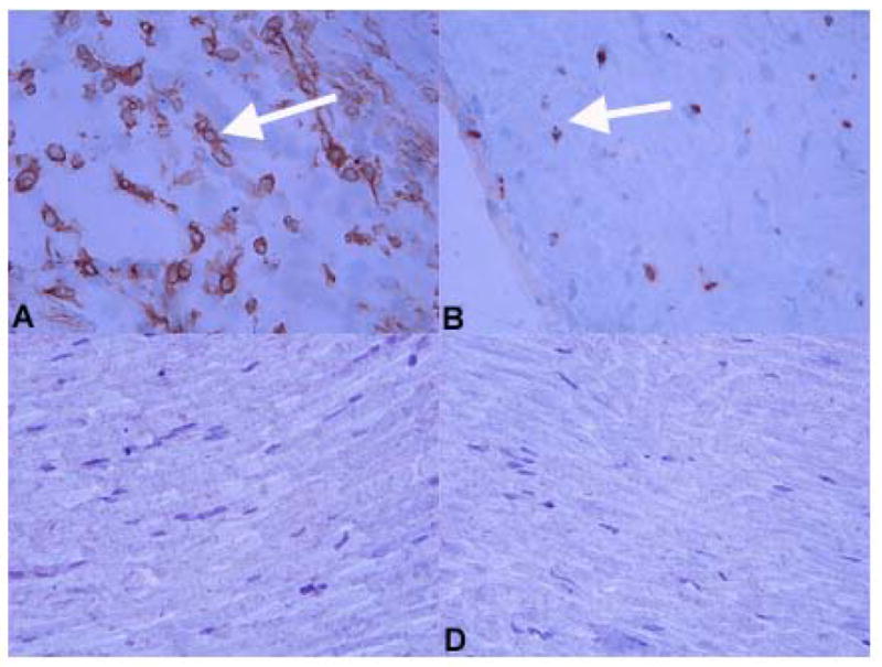
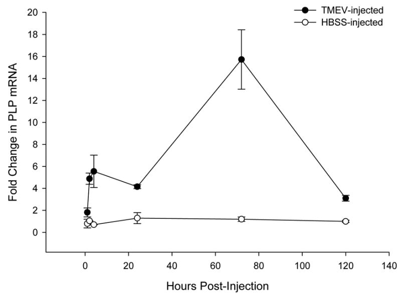
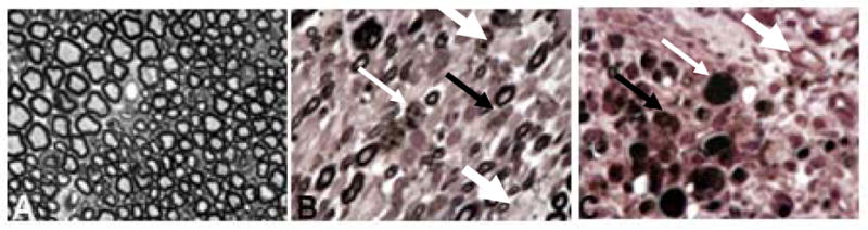
Demyelination of the human peripheral nervous system (PNS) can be caused by diverse mechanisms including viral infection. Despite association of several viruses with the development of peripheral demyelination, animal models of the condition have been limited to disease that is either autoimmune or genetic in origin. We describe here a model of PNS demyelination based on direct injection of sciatic nerves of mice with the cardiovirus, Theiler's murine encephalomyelitis virus (TMEV). Sciatic nerves of FVB mice develop inflammatory cell infiltration following TMEV injection. Schwann cells and macrophages are infected with TMEV. Viral replication is observed initially in the sciatic nerves and subsequently the spinal cord. Sciatic nerves are demyelinated by day 5 post-inoculation (p.i.). Injecting sciatic nerves of scid mice resulted in increased levels of virus recovered from the sciatic nerve and spinal cord relative to FVB mice. Demyelination also occurred in scid mice and by 12 days p.i., hindlimbs were paralyzed. This new model of virus-induced peripheral demyelination may be used to dissect processes involved in protection of the PNS from viral insult and to study the early phases of lesion development.


Figures

Figure 1
Figure 1. Immunolocalization of TMEV antigens in the sciatic nerve of FVB mice
Virus antigen positive cells are apparent in sciatic nerves at 3 days post-infection of the sciatic nerve with TMEV; staining of a TMEV-injected sciatic nerve is shown in (A). Dark reaction product indicates positive staining (examples shown by white arrowhead). No immunoreactivity was observed in the sciatic nerve of an HBSS-injected mouse (B). Following injection of UV-inactivated virus into the sciatic nerve, viral proteins were not detected by immunohistochemistry (C). Slides were stained using the ABC Methodology as described previously (Drescher, Nguyen et al., 1998). Development was carried out using DAB. Double immunofluorescent staining was performed using markers specific for myelinating cells (D, F, G) combined with identification of TMEV (E) as described in the Methods. Single channel images are shown (D, E) as well as merged images (F, G). Staining is shown for sciatic nerves from FVB mice at 5 days post-inoculation (D–F). A merge image of the uninfected contralateral sciatic nerve is negative for TMEV (G). Controls included staining a normal mouse brain with the antibody to myelinating cells to demonstrate specificity for white matter areas of the brain (H). A DAPI image of the brain is shown for comparison purposes (I). Original magnification X480 (A–C), X1000 (D–G), X80 (H, I).
Figure 2
Figure 2. Immunostaining for immune cells in the sciatic nerves of virus (A, B) and control (C, D) injected mice
At day 4 p.i., macrophages (A) and T cells (B) were detected in sciatic nerves of virus-injected FVB mice (examples shown by white arrowhead). Neither macrophages (C) or T cells (D) were identified in the sciatic nerve of control, HBSS-injected female FVB mice. Markers used to identify these cell types were F4/80 (macrophages) and CD3 (T cells). Slides were stained using the ABC immunoperoxidase technique as described (Methods). Development was carried out using DAB. Dark colored reaction product indicates positive staining. Original magnification X480.
Figure 3
Figure 3. Real-time RT- PCR demonstrates that PLP mRNA levels peak at day 3 post-injection of the sciatic nerve with TMEV (solid circles)
A 4.9 ± 0.5 -fold relative increase in PLP mRNA levels is evident as early as 2 hours after sciatic nerve injection with TMEV (solid circles; p = 0.02). By day 3 p.i. PLP transcript levels peak, with a 15.7 ± 2.7-fold increase in mRNA levels as compared to uninjected control sciatic nerves (p < 0.032). This increase in PLP mRNA levels begins to subside by day 5 p.i. with virus, but still remain 3.1 ± 0.3-fold higher than baseline levels (p = 0.02). Additional control mice were injected with HBSS (open circles) into the sciatic nerve. No significant alterations in PLP mRNA levels were observed over 5 days. Standardization was performed using GAPDH as a housekeeping gene. Real-time PCR was performed using TaqMan methodology (Methods) on an ABI 7000 SDS Sequence Detection System (Applied Biosystems).
Figure 4
Figure 4. Pathology of the sciatic nerve of virus-injected (B,C) and HBSS-injected (A) mice
HBSS-injected sciatic nerves are characterized by uniform, well-preserved myelin around the axons; infiltrating cells are absent (A). Mice injected with TMEV (B, C) show demyelination of the axons (demyelination presents as paler circles versus the dark well-myelinated axons; thick white arrow). In addition, the sciatic nerve contains cells which, due to their foamy appearance, appear to be macrophages (thin, white arrow) as well as myelin debris (thin black arrow). Sciatic nerves were osmicated, embedded in plastic and 1 micron sections prepared. Slides were stained with 4% para-phenylenediamene (PPD) and examined by light microscopy. Original magnification X1200.

Similar articles

Cited by

References

    1. Bernhardt G, Bibb JA, Bradley J, Wimmer E. Molecular characterization of the cellular receptor for poliovirus. Virol. 1994;199:105–113. - PubMed
    1. Bitan M, Or R, Shapira MY, Mador N, Resnick IB, Saleh N, Weinberger KM, Samuel S, Schechter E, Slavin S, Wolf DG. Early-onset Guillain-Barre syndrome associated with reactivation of Epstein-Barr virus infection after nonmyeloablative stem cell transplantation. Clin Infect Dis. 2004;39:1076–1078. - PubMed
    1. Bosma GC, Custer RP, Bosma MJ. A severe combined immunodeficiency mutation in the mouse. Nature. 1983;301:527–530. - PubMed
    1. Bour-Jordan H, Thompson HL, Bluestone JA. Distinct effector mechanisms in the development of autoimmune neuropathy versus diabetes in nonobese diabetic mice. J Immunol. 2005;175:5649–5655. - PubMed
    1. Brannagan TH, 3, Zhou Y. HIV-associated Guillain-Barre syndrome. J Neurol Sci. 2003;208:39–42. - PubMed

Publication types

MeSH terms