. 2006;34(19):5603-12.
doi: 10.1093/nar/gkl619. Epub 2006 Oct 5.

Expression of C-terminal deleted p53 isoforms in neuroblastoma

Affiliations

Expression of C-terminal deleted p53 isoforms in neuroblastoma

David Goldschneider et al. Nucleic Acids Res. 2006.

Abstract

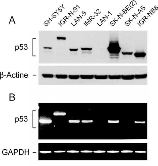
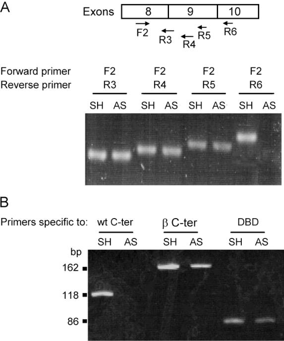
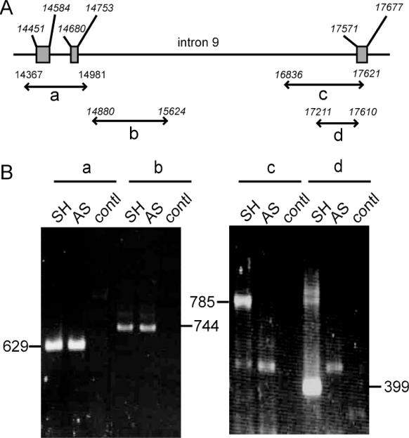
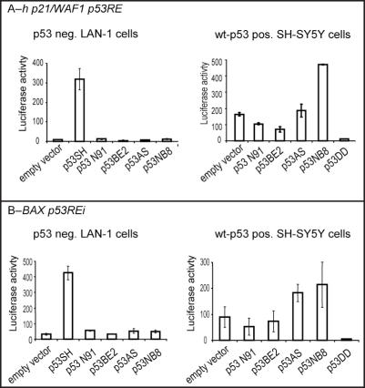
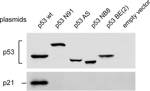
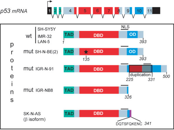
The tumor suppressor gene, p53, is rarely mutated in neuroblastomas (NB) at the time of diagnosis, but its dysfunction could result from a nonfunctional conformation or cytoplasmic sequestration of the wild-type p53 protein. However, p53 mutation, when it occurs, is found in NB tumors with drug resistance acquired over the course of chemotherapy. As yet, no study has been devoted to the function of the specific p53 mutants identified in NB cells. This study includes characterization and functional analysis of p53 expressed in eight cell lines: three wild-type cell lines and five cell lines harboring mutations. We identified two transcription-inactive p53 variants truncated in the C-terminus, one of which corresponded to the p53beta isoform recently identified in normal tissue by Bourdon et al. [J. C. Bourdon, K. Fernandes, F. Murray-Zmijewski, G. Liu, A. Diot, D. P. Xirodimas, M. K. Saville and D. P. Lane (2005) Genes Dev., 19, 2122-2137]. Our results show, for the first time, that the p53beta isoform is the only p53 species to be endogenously expressed in the human NB cell line SK-N-AS, suggesting that the C-terminus truncated p53 isoforms may play an important role in NB tumor development.


Figures

Figure 1
Figure 1
p53 expression of different neuroblastoma cell lines. (A) Western blot from protein total extracts (upper panel); β-actin was used as loading control (lower panel). (B) RT–PCR using F1-R7 primers (Table 1); amplified fragment was normalized by GAPDH.
Figure 2
Figure 2
Detection of p53 mRNA abnormalities in SK-N-AS (AS) in comparison with SH-SY5Y (SH) cells. (A) Amplification of p53 cDNA using primers from exons 8 to 10 as indicated below each arrow; for precise position see Table 1 and GenBank K03199: F2 (forward primer in exon 8); R3 (reverse primer at the junction of exon 8/9); R4 (the first moiety of exon 9); R5 (exon 9, 30 nt downstream R4); R6 (beginning of exon 10). Note that no amplification was observed in SK-N-AS from exon 10 (last lane), in contrast to SH-SY5Y. (B) RT–PCR from SK-N-AS compared to SH-SY5Y cells. Specific primers (Table 1) were used to amplify the DBD, the p53β isoform (β) and the C-terminal domain (C-ter).
Figure 3
Figure 3
Identification of a deletion spanning the intron9/exon 10 junction of SK-N-AS p53 gene. (A) Schematic representation of p53 gene from intron 7 to intron 10 with the position of amplified fragments; (B) PCR fragments amplified from SH-SY5Y (SH) and SK-N-AS (AS) DNA with the primer pairs a, b, c and d. The primer sequences are given in Table 1; contl: PCR performed in parallel without DNA.
Figure 4
Figure 4
p53 transactivation ability using yeast-based assay (FASAY). (A) Schematic representation of the analysis of p53 mutants using the yeast homologous recombination expression vector pRDI-22 carrying the 5′ and 3′ ends of the p53 open reading frame and the split forms, pFW35 and pFW34 (lacking p53 fragment from amino acids 66 to 210 for split 5′ and 211–348 for split 3′, respectively) transfected into YPH500 Ade2 yeast strain. This strain repairs double-strand breaks in transfected plasmids by homologous recombination as ‘gap repair’ (see text). (B) Photographs of yeast colonies showing 100% wild-type p53 where all colonies are white (a), or special mutated p53 by duplication of exons 7–9 found in IGR-N-91 cells (b), where white and red colonies were mixed (see also Table 2), and mutated p53 such as those in SK-N-BE(2), where all colonies are red (c).
Figure 5
Figure 5
p53 transactivation ability by luciferase test using plasmid pE1B-hWAF1(A) and Bax (B). p53-deficient LAN-1 cells (p53-) (left panel) or SH-SY5Y (p53+) (right panel) were cotransfected with 0.5 μg of the luciferase reporter gene containing the human p21/WAF1-p53-responsive element and with 1 μg of the expressing vector as indicated. Cells were collected and subject to luciferase assay, 24 h following cotransfection. The values represent mean relative luciferase activity from three independent experiments. SH-SY5Y, IGR-N-91, SK-N-BE(2), SK-N-AS and IGR-NB8 were termed as SH, N91, BE(2), AS and NB8, respectively.
Figure 6
Figure 6
Western blot showing induction of p21/WAF1 protein by plasmid-recombinant expression vector of p53 variant transfected into p53-negative LAN-1 cells. LAN-1 cells were seeded onto 6-well plates. At a density of ∼60%, confluence cells were transfected with recombinant vector using Lipofectamine 2000 reagent according to the supplier's instructions (Invitrogen). To ascertain the transfection efficiency, cells were transfected in parallel experiments with pEGFP-C1 vector (Promega). The empty vector was used as a control. As shown in Figure 1, note that the p53 protein from the IGR-N-91 cells analyzed by SDS–PAGE migrated more slowly than the wild-type p53 due to duplication of exons 7-8-9 as described previously (13).
Figure 7
Figure 7
Western blot showing induction of endogenous p21/WAF1 in different neuroblastoma cell lines in response to 10 μM cisplatin treatment. Protein p21/WAF1 induction was observed only in the three wild-type p53 NB cell lines and the protein was differentially accumulated.
Figure 8
Figure 8
Visualization of chromosome 17 copy number and p53 genes by FISH experiments. Chromosome 17 centromere probe is green labeled and TP53 gene probe red labeled. Only the predominant clone of each cell line is presented and its frequency indicated.
Figure 9
Figure 9
Structure of p53 proteins in different neuroblastoma cell lines. The three functional domains are represented: TAD, transactivation domain; DBD, DNA-binding domain; OD, oligomerization domain. The wild-type p53 gene in SH-SY5Y, IMR-32 and LAN-5 cells contains 11 exons that encode 393 amino acids. In SK-N-BE(2) cells, p53 is mutated at codon 135 (*), which converts cysteine to phenylalanine. In IGR-N-91 cells, a duplication of exons 7-8-9 adds an additional 107 amino acids leading to a total of 500. In SK-N-AS cells, a mutation due to alternate splicing downstream of exon 9 leads to a protein of 341 amino acids whereas in IGR-NB8 cells, the p53 protein ends at 326 amino acids owing to the mutation E326STOP.

References

    1. Hainaut P., Hollstein M. p53 and human cancer: the first ten thousand mutations. Adv. Cancer Res. 2000;77:81–137. - PubMed
    1. Soussi T., Beroud C. Assessing TP53 status in human tumours to evaluate clinical outcome. Nature Rev. Cancer. 2001;1:233–240. - PubMed
    1. Beroud C., Soussi T. The UMD-p53 database: new mutations and analysis tools. Hum. Mutat. 2003;21:176–181. - PubMed
    1. Soussi T., Kato S., Levy P.P., Ishioka C. Reassessment of the TP53 mutation database in human disease by data mining with a library of TP53 missense mutations. Hum. Mutat. 2005;25:6–17. - PubMed
    1. Moll U.M., Laquaglia M., Benard J., Riou G. Wild-type p53 protein undergoes cytoplasmic sequestration in undifferentiated neuroblastomas but not in differentiated tumors. Proc. Natl Acad. Sci. USA. 1995;92:4407–4411. - PMC - PubMed

Publication types

Substances

Associated data