. 2007 Jan 1;92(1):225-33.
doi: 10.1529/biophysj.106.091561. Epub 2006 Oct 6.

Contour length and refolding rate of a small protein controlled by engineered disulfide bonds

Affiliations

Contour length and refolding rate of a small protein controlled by engineered disulfide bonds

Sri Rama Koti Ainavarapu et al. Biophys J. .

Abstract

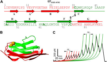
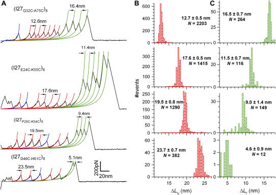
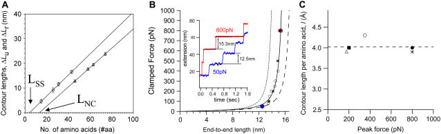
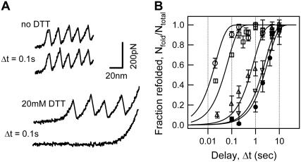
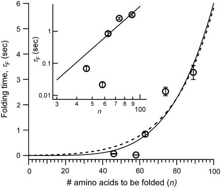
The introduction of disulfide bonds into proteins creates additional mechanical barriers and limits the unfolded contour length (i.e., the maximal extension) measured by single-molecule force spectroscopy. Here, we engineer single disulfide bonds into four different locations of the human cardiac titin module (I27) to control the contour length while keeping the distance to the transition state unchanged. This enables the study of several biologically important parameters. First, we are able to precisely determine the end-to-end length of the transition state before unfolding (53 Angstrom), which is longer than the end-to-end length of the protein obtained from NMR spectroscopy (43 Angstrom). Second, the measured contour length per amino acid from five different methods (4.0 +/- 0.2 Angstrom) is longer than the end-to-end length obtained from the crystal structure (3.6 Angstrom). Our measurement of the contour length takes into account all the internal degrees of freedom of the polypeptide chain, whereas crystallography measures the end-to-end length within the "frozen" protein structure. Furthermore, the control of contour length and therefore the number of amino acids unraveled before reaching the disulfide bond (n) facilitates the test of the chain length dependence on the folding time (tau(F)). We find that both a power law scaling tau(F) lambda n(lambda) with lambda = 4.4, and an exponential scaling with n(0.6) fit the data range, in support of different protein-folding scenarios.


Figures

FIGURE 1
FIGURE 1
Design and reduction of individual disulfide bonds engineered into a titin immunoglobulin module (I27). Amino acid sequence (A) and cartoon representation (B) of the I27G32C-A75C protein. The oxidized disulfide bond (B; golden-yellow spheres) “traps” 43 amino acids encompassing the C, D, E, and F β-strands (green), whereas the A, A', B, and G β-strands (red) remain “unsequestered”. (C) Force-extension curve obtained by pulling an (I27G32C-A75C)8 polyprotein in the presence of 100 mM DTT. The trace shows two distinct, equally spaced sawtooth patterns, which appear in sequence. Each peak in the first sawtooth pattern corresponds to the unsequestered unfolding of a single I27 module up to the disulfide bond. The second sawtooth observed at a higher force corresponds to the sequential reduction of individual disulfide bonds in each module. The WLC model fits are shown in thick lines, and the contour length per module for the unsequestered unfolding (ΔLu) and disulfide bond reduction (ΔLr) are 12.6 nm and 16.4 nm, respectively.
FIGURE 2
FIGURE 2
Contour length increments depend on the position of the engineered disulfide bond. (A) Force-extension curves in the presence of DTT were obtained from four different polyprotein constructs: (I27G32C-A75C)8, (I27E24C-K55C)8, (I27P28C-K54C)8, and (I27D46C-H61C)8. Each trace is fit by the WLC of polymer elasticity. The WLC fits measure the contour length increments of unfolding (red lines; ΔLu) and of the bond reduction events (green lines; ΔLr). Fits shown in blue indicate the unfolding of modules where the disulfide bond was reduced in solution before mechanical unfolding. The contour length increase of full-length I27 (ΔLf) ≈ 28.4 nm is equivalent to the contour length observed on unfolding wild-type I27 (7). (B) Histograms of the measured values of ΔLu for each construct. (C) Histograms of the measured values of ΔLr for each construct. These values are summarized in Table 1. Note that for each construct ΔLu + ΔLr ≈ ΔLf, demonstrating that the end-to-end length of the transition state remains the same for all the constructs.
FIGURE 3
FIGURE 3
Distance to the transition state is unperturbed on disulfide bond insertion into I27. The unfolding forces of the (I27E24C-K55C)8 protein were obtained by varying the pulling speed from 40 nm/s to 4000 nm/s (solid circles). The solid line corresponds to the Monte Carlo simulation where the distance to the transition state is Δxu = 0.25 nm, and the spontaneous unfolding rate constant is formula image. The value of Δxu is identical to that of the wild-type I27 (0.25 nm), indicating that the position of the transition state remains the same. However, the spontaneous unfolding rate constant is increased by 5.6 times compared to that of the wild-type I27 (3.3 × 10⁻⁴ −1).
FIGURE 4
FIGURE 4
Determination of the contour length increment per amino acid (l). (A) Plot of the contour lengths ΔLu (triangles) and ΔLr (circles) versus the corresponding number of unsequestered and trapped amino acids (see Table 1) for all four polyprotein constructs. The slope of a linear fit to the data (solid lines) accurately measures the contour length per amino acid obtained by extending the unsequestered (lu = 3.9 Å/aa) and trapped (lr = 4.3 Å/aa) amino acids. The extrapolated intercepts with the abscissa (LNC = 13.4 aa and LSS = 4.3 aa) indicate the end-to-end length of the transition state of unfolding and the distance between the Cα atoms of the disulfide-bonded cysteines in the fully extended form. (B) Force-clamp experiments stretch the molecule under a constant force and measure the end-to-end length of 43 amino acids released by the forced reduction of the disulfide bond in the (I27G32C-A75C)8 polyprotein. Protein extensions measured at two clamping forces, 50 pN and 800 pN, are shown in the inset, giving rise to step sizes of 12.5 nm and 15.3 nm, respectively. A plot of the average step sizes (open circles and solid circles) as a function of force is fit with the WLC theory, using the contour length Lr = 16.4 nm (lr = 4.0 Å/aa) and the persistence length of 0.3 nm. We superimpose four points (×) obtained by SMD simulations (rescaled by the protein length) to show further agreement with the obtained experimental values. WLC curves created using the contour lengths corresponding to the crystalline (3.6 Å/aa) and the linear (4.3 Å/aa) end-to-end lengths of an amino acid are shown as dotted and dashed lines, respectively, failing to fit the experimental data. (C) Contour length per amino acid (l) as a function of peak force measured in force-extension experiments of unsequestered unfolding (triangle), disulfide bond reduction (circle), unfolding of Gly5 inserted I27 (solid square), force-clamp experiments (solid circle), and SMD simulations (*). The dotted line represents the average (4.02 Å/aa) of l obtained using these five methods.
FIGURE 5
FIGURE 5
Folding time dependence on the chain length (n, number of amino acids). (A) Force-extension curves of the (I27E24C-K55C)8 polyprotein unfolding and refolding cycles. The top two recordings were obtained with a relaxation delay of 0.1 s in the absence of DTT (top trace) and in 20 mM DTT (bottom trace). The same number of peaks in the consecutive unfolding trajectories provides evidence that the protein with an oxidized disulfide bond completely refolded in 0.1 s, whereas the absence of peaks in the second unfolding trace in DTT shows that the reduced protein is yet to begin folding at 0.1 s. (B) Graph of the fraction of refolded modules (Nfold/Ntotal) versus the delay time (Δt). Data obtained from (I27G32C-A75C)8 (squares), (I27E24C-K55C)8 (circles), (I27P28C-K54C)8 (triangles), and (I27D46C-H61C)8 (inverted triangles), as well as the reduced protein (I27E24C-K55C)8 obtained in 20 mM DTT (solid circles) are shown. The solid lines are fits of the data to the function P(t) = 1 − exp(−t/τF).
FIGURE 6
FIGURE 6
Plot of folding time (τF) versus the number of unsequestered amino acids (n). The solid lines show the power law fit, τF = B nλ, where B = 10−8 s, λ = 4.4 (solid line), and the fit to a barrier-activated process, τF = A exp(Knγ), where A = 0.002 s, K = 0.6, and γ = 0.57 (dotted line). Inset shows the log-log plot of folding times as a function of the chain length, where the power law fit is a straight line.

References

    1. Bustamante, C., J. F. Marko, E. D. Siggia, and S. Smith. 1994. Entropic elasticity of lambda-phage DNA. Science. 265:1599–1600. - PubMed
    1. Yang, G., C. Cecconi, W. A. Baase, I. R. Vetter, W. A. Breyer, J. A. Haack, B. W. Matthews, F. W. Dahlquist, and C. Bustamante. 2000. Solid-state synthesis and mechanical unfolding of polymers of T4 lysozyme. Proc. Natl. Acad. Sci. USA. 97:139–144. - PMC - PubMed
    1. Dietz, H., and M. Rief. 2006. Protein structure by mechanical triangulation. Proc. Natl. Acad. Sci. USA. 103:1244–1247. - PMC - PubMed
    1. Erickson, H. P. 1994. Reversible unfolding of fibronectin type III and immunoglobulin domains provides the structural basis for stretch and elasticity of titin and fibronectin. Proc. Natl. Acad. Sci. USA. 91:10114–10118. - PMC - PubMed
    1. Trombitas, K., M. Greaser, S. Labeit, J. P. Jin, M. Kellermayer, M. Helmes, and H. Granzier. 1998. Titin extensibility in situ: entropic elasticity of permanently folded and permanently unfolded molecular segments. J. Cell Biol. 140:853–859. - PMC - PubMed

Publication types

LinkOut - more resources