. 2006 Oct 17;103(42):15529-34.
doi: 10.1073/pnas.0607262103. Epub 2006 Oct 6.

Critical role for the beta regulatory subunits of Cav channels in T lymphocyte function

Affiliations

Critical role for the beta regulatory subunits of Cav channels in T lymphocyte function

Abdallah Badou et al. Proc Natl Acad Sci U S A. .

Abstract

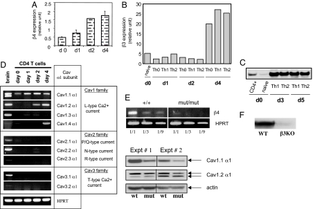
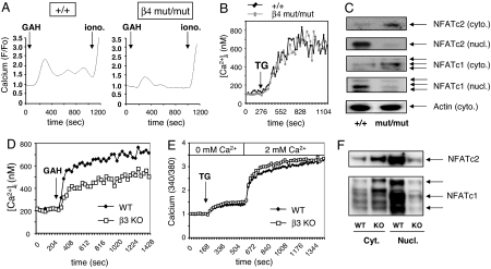
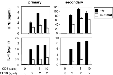
Calcium ion is a universal signaling intermediate, which is known to control various biological processes. In excitable cells, voltage-gated calcium channels (Cav) are the major route of calcium entry and regulate multiple functions such as contraction, neurotransmitter release, and gene transcription. Here we show that T lymphocytes, which are nonexcitable cells, express both regulatory beta and pore-forming Cav1 alpha1 subunits of Cav channels, and we provide genetic evidence for a critical role of the Cav beta3 and Cav beta4 regulatory subunits in T lymphocyte function. Cav beta-deficient T lymphocytes fail to acquire normal functions, and they display impairment in the T cell receptor-mediated calcium response, nuclear factor of activated T cells activation, and cytokine production. In addition, unlike in excitable cells, our data suggest a minimal physiological role for depolarization in Cav channel opening in T cells. T cell receptor stimulation induces only a small depolarization of T cells, and artificial depolarization of T cells using KCl does not lead to calcium entry. These observations suggest that the Cav channels expressed by T cells have adopted novel regulation/gating mechanisms.


Conflict of interest statement

The authors declare no conflict of interest.

Figures

Fig. 1.
Fig. 1.
Expression of Cav channel subunits by CD4 T cells. Purified total CD4 T cells (CD4+) or naïve T cells (CD62L-high and CD44-low) were left unstimulated (day 0) or were stimulated with plate-bound anti-CD3 plus anti-CD28 Abs for the indicated period. (A and B) Analysis of β4 and β3 subunit mRNA expression by CD4 T cells using real-time quantitative PCR. (C) Western blot analysis of β3 subunit expression in CD4 T cells. (D) Expression of the Cav channel α1 subunits by CD4 T cells at different stages of differentiation. (E) Expression of Cav channel subunits by β4 mutant T cells assessed by using RT-PCR and Western blot. (F) Western blot analysis of the expression of β3 subunit in effector WT or β3 KO T cells. Results in A and D are representative of at least three independent experiments. For B, C, E, and F, two experiments were performed.
Fig. 2.
Fig. 2.
Calcium/NFAT pathway is impaired in both β4- and β3-deficient CD4 T cells. (A and D) The calcium response of CD4 T cells from β4-deficient (mut/mut) or β3-deficient (β3 KO) mice and WT littermate was evaluated by using 2C11 anti-CD3 Ab and goat anti-hamster (GAH) in a cross-linking system. (B and E) Calcium response of WT and β4- or β3-deficient CD4 T lymphocytes upon stimulation with thapsigargin (1 μM) under physiological Ca2+ concentrations (B) or in Ca2+ add-back experiments (E). (C) Western blot assay was performed on cytoplasmic (cyto.) and nuclear (nucl.) extracts prepared from β4 mutant CD4 T cells and control littermate stimulated for 48 h in the presence of human IL-2. Actin was used as internal control. (F) Analysis of β3-deficient CD4 T cells for NFAT nuclear translocation, using Western blot, after 18 h of TCR stimulation. Results are representative of five (A and D), three (B, C, and E), and two (F) independent experiments.
Fig. 3.
Fig. 3.
Defective CD4 T cell differentiation in the absence of functional β4 subunit. CD4 T cells were stimulated by plate-bound anti-CD3 plus anti-CD28. Three days later, IFNγ and IL-4 production was measured by ELISA (primary). In some experiments cells were stimulated for 4 days, then washed and restimulated (secondary) by plate-bound anti-CD3 Ab. Twenty-four hours later, IFNγ and IL-4 production was measured by ELISA. Results are representative of at least three independent experiments.
Fig. 4.
Fig. 4.
TCR stimulation induces only a small depolarization, and depolarization by KCl does not trigger T cell Cav opening. (A Upper) A modest depolarization of T lymphocytes after TCR stimulation (in black). Depolarization was assessed with the voltage-sensitive dye DiBAC4 (3) by using flow cytometric analysis. (A Lower) Depolarization using different concentrations of KCl (10–40 mM). Results shown in A are representative of four independent experiments. (B) CD4 T cells were purified from C57BL/6 mice and then differentiated under Th1 (IL-12 plus anti-IL-4), Th2 (IL-4 plus anti-IFNγ), or Th0 (no cytokine) conditions using Abs to CD3 and CD28. The calcium response of fura-2-loaded T or C2C12 cells was evaluated after the addition of KCl (40 mM). Results shown in B are representative of three independent experiments.

References

    1. Cantrell D. Annu Rev Immunol. 1996;14:259–274. - PubMed
    1. Lewis RS. Annu Rev Immunol. 2001;19:497–521. - PubMed
    1. Zweifach A, Lewis RS. Proc Natl Acad Sci USA. 1993;90:6295–6299. - PMC - PubMed
    1. Feske S, Gwack Y, Prakriya M, Srikanth S, Puppel SH, Tanasa B, Hogan PG, Lewis RS, Daly M, Rao A. Nature. 2006;441:179–185. - PubMed
    1. Vig M, Peinelt C, Beck A, Koomoa DL, Rabah D, Koblan-Huberson M, Kraft S, Turner H, Fleig A, Penner R, Kinet JP. Science. 2006;312:1220–1223. - PMC - PubMed

Publication types