. 2006 Oct;18(10):2792-806.
doi: 10.1105/tpc.106.044016. Epub 2006 Oct 6.

The tomato NBARC-LRR protein Prf interacts with Pto kinase in vivo to regulate specific plant immunity

Affiliations

The tomato NBARC-LRR protein Prf interacts with Pto kinase in vivo to regulate specific plant immunity

Tatiana S Mucyn et al. Plant Cell. 2006 Oct.

Abstract

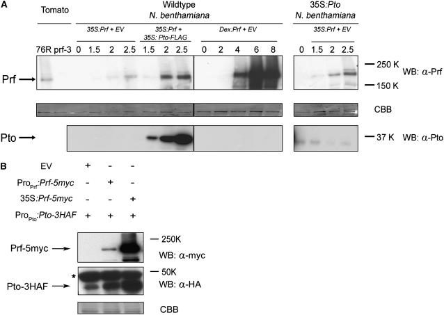
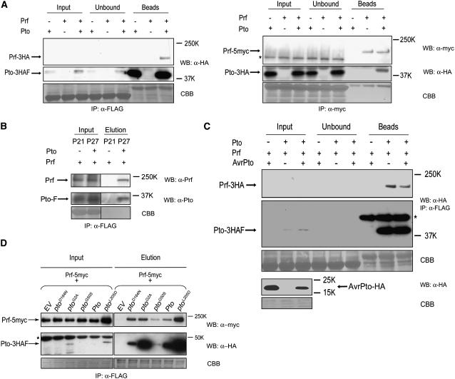
Immunity in tomato (Solanum lycopersicum) to Pseudomonas syringae bacteria expressing the effector proteins AvrPto and AvrPtoB requires both Pto kinase and the NBARC-LRR (for nucleotide binding domain shared by Apaf-1, certain R gene products, and CED-4 fused to C-terminal leucine-rich repeats) protein Prf. Pto plays a direct role in effector recognition within the host cytoplasm, but the role of Prf is unknown. We show that Pto and Prf are coincident in the signal transduction pathway that controls ligand-independent signaling. Pto and Prf associate in a coregulatory interaction that requires Pto kinase activity and N-myristoylation for signaling. Pto interacts with a unique Prf N-terminal domain outside of the NBARC-LRR domain and resides in a high molecular weight recognition complex dependent on the presence of Prf. In this complex, both Pto and Prf contribute to specific recognition of AvrPtoB. The data suggest that the role of Pto is confined to the regulation of Prf and that the bacterial effectors have evolved to target this coregulatory molecular switch.


Figures

Figure 1.
Figure 1.
Pto and Prf Are Coincident in a Signal Transduction Pathway Leading to Disease Resistance. (A) Requirement of Pto for constitutive disease resistance in tomato mediated by Prf overexpression. Left panel, growth of Pst strain T1 on tomato genotypes as indicated. The values are means of three samples, from three different plants, treated equivalently in the same experiment. Error bars indicate sd. The graph presented is representative of three replicate experiments. Right panel, RNA gel blot showing Prf expression. The 18S rRNA band is shown as a loading control. (B) Codependence of Prf and Pto for the overexpression HR in N. benthamiana. Genes expressed from the cauliflower mosaic virus 35S and ProPto promoters are indicated, and EV indicates the empty binary vector control. The photograph was taken 3.5 d after infiltration. (C) The Prf overexpression HR requires normal Pto signaling capabilities. Left panel, 35S:Prf was coexpressed with wild-type Pto, the kinase-knockout mutant ptoD164N, the N-myristoylation mutant ptoG2A, or the weak kinase variant ptoG50S. The photograph was taken 5 d after infiltration. Right panel, protein accumulation was confirmed by protein gel blot using the anti-HA antibody for Prf-3HA detection (top gel) or polyclonal anti-Pto antiserum (middle gel). wt indicates untransformed tissue, and other constructs are as indicated. Coomassie blue (CBB) staining of the protein gel blot membrane confirmed equivalent protein loading (bottom gel).
Figure 2.
Figure 2.
Prf Is a Signaling Protein Regulated by Pto. (A) Overexpression of Prf from a DEX-inducible promoter (Dex:Prf) induces a Pto-independent HR. Prf expression was induced (+DEX) or mock-induced (−DEX) 1.5 d after infiltration. The photographs were taken 24 h after induction. (B) The Dex:Prf HR is independent of Nb Pth1. Left panel, transient expression of Dex:Prf (left) or 35S:AvrPto-GFP (right) in wild-type N. benthamiana containing an empty VIGS construct (top) or silenced for Nb Pth1 (bottom). The photographs were taken 2.5 d after infiltration, and gene expression was induced as for (A). Right panel, AvrPto-GFP expression in silenced plants was confirmed by protein gel blot using an anti-GFP antibody. (C) Key pto mutants dominantly suppress the Dex:Prf HR. Dex:Prf was coexpressed in N. benthamiana leaves (top panel) with various 35S:pto mutants as indicated or an EV control. The leaf was induced with DEX 1.5 d after infiltration and photographed 24 h after induction. Leaf tissues were harvested for protein extraction 6 h after induction. Accumulation of Pto and Prf proteins was confirmed by protein gel blot as indicated, and equal protein loading was confirmed by Coomassie Brilliant Blue (CBB) staining of the protein gel blot membrane. The asterisk indicates that the tissue displayed HR at the time of harvest.
Figure 3.
Figure 3.
Pto and Prf Contribute Reciprocally to Protein Accumulation. (A) Pto contributes to Prf accumulation in planta. Prf was detected from protein extracts of RioGrande 76R or prf3 tomato or from wild-type and 35S:Pto transgenic N. benthamiana plants (Rommens et al., 1995). Left, extracts of tomato containing (76R) or lacking (prf3) a functional Prf gene. Middle, expression of 35S:Prf or Dex:Prf in wild-type N. benthamiana, with (35S:Pto) or without (EV) Pto as indicated. For 35S:Prf expression, numbers refer to days after infiltration with Agrobacterium, whereas for Dex:Prf, numbers refer to hours after induction with DEX. Right, expression of 35S:Prf in stable transgenic N. benthamiana plants expressing 35S:Pto (38-12). Note the low level of Pto accumulation in these plants. Proteins were detected specifically as indicated, and equal protein loading was confirmed by Coomassie Brilliant Blue (CBB) staining of the protein gel blot membrane. (B) Prf contributes to Pto accumulation. ProPto:Pto-3HAF was coexpressed with EV, ProPrf:Prf-5myc, or 35S:Prf-5myc in N. benthamiana leaves. Tissues were harvested at 2.5 d after infiltration. Equal amounts of protein derived from crude extracts of infiltrated tissues were loaded on SDS-PAGE gels for immunoblotting. Pto and Prf were detected using antisera as indicated. The asterisk indicates a nonspecific cross-reacting band.
Figure 4.
Figure 4.
Prf Interacts with Pto in Vivo. (A) Prf and Pto coimmunoprecipitate from N. benthamiana extracts. Left, N. benthamiana leaves were transiently transformed with ProPto:Pto-HAF, ProPrf:Prf-3HA, or a 1:1 mixture of these strains. Strains for individual gene expression were balanced with A. tumefaciens containing an empty vector construct. Infiltrated leaves were harvested 3 d after infiltration for extraction of proteins, and protein extracts were subjected to immunoprecipitation as indicated. Equal amounts by volume of crude extract (Input) and supernatant after immunoprecipitation (Unbound) were loaded onto the SDS-PAGE gel for immunoblotting. The bead fractions (Beads) were ∼80 times more concentrated than the crude extract. Right, N. benthamiana leaves transiently transformed as above with ProPto:Pto-3HA, ProPrf:Prf-5myc, or a 1:1 mixture of these strains. Samples were prepared as above, except for the use of anti-myc agarose beads for immunoprecipitation. The bead fractions were ∼20 times more concentrated than the crude extract. Prf-5myc was undetectable in the crude extract. The asterisk indicates a cross-reacting band. Equal protein loading was confirmed by Coomassie blue (CBB) staining of the protein gel blot membrane. (B) Prf coimmunoprecipitates with Pto in tomato extracts. Protein extracts from transgenic tomato lines expressing ProPto:Pto-FLAG (P27) or EV (P21) were subjected to immunoprecipitation using anti-FLAG beads before specific elution of bound proteins with the FLAG peptide (Elution). (C) The Pto–Prf interaction is not disrupted by AvrPto. Transgenic Dex:avrPto-HA N. benthamiana plants were transiently transformed with ProPto:Pto-3HAF, ProPrf:Prf-3HA, or a 1:1 mixture of these strains as above. Transformed leaves were treated with DEX 2.5 d after infiltration to induce AvrPto expression. Tissues were harvested before treatment or 6 h after treatment with DEX. Protein extracts were immunoprecipitated with anti-FLAG beads followed by immunoblotting with anti-HA antibody to detect Prf-3HA, AvrPto-HA, and Pto-3HAF. The bead fractions were ∼80 times more concentrated than the crude extract in this experiment. The asterisk indicates the heavy chain of the anti-FLAG antibody released from the beads. (D) Interaction of Prf with key pto mutants. N. benthamiana plants were first transiently transformed with 35S:Prf-5myc, then infiltrated 24 h later with EV, ProPto:ptoD164N-3HAF, ProPto:ptoG2A-3HAF, ProPto:ptoG50S-3HAF, ProPto:Pto-3HAF, or ProPto:ptoL205D-3HAF. Tissues were harvested 2 d after the second infiltration. Protein extracts were subjected to immunoprecipitation using anti-FLAG beads before elution of bound proteins with the FLAG peptide. Equivalent amounts of crude extract before immunoprecipitation (Input) or the final eluates (Elution) were loaded onto SDS-PAGE gels for immunoblotting. Pto and Prf were detected using specific antisera as indicated. The asterisk indicates a cross-reacting band.
Figure 5.
Figure 5.
Prf and Pto Coelute in a High Molecular Weight Fraction during SEC. (A) Elution of Prf by SEC–fast protein liquid chromatography (FPLC). Total protein extracts from wild-type 76R (Prf/Prf) (top panel) or prf3 (prf3/prf3) (bottom panel) tomato leaves were separated by SEC-FPLC before concentration of the fractions preceding SDS-PAGE and protein gel blotting with specific antisera as indicated. Numbers refer to the volume of elution (in mL) beyond the void volume of the column. The peaks of elution of the molecular weight standards are indicated. (B) Elution of Prf and Pto by SEC-FPLC after transient expression in N. benthamiana. 35S:Prf was coexpressed with 35S:Pto-3HAF in leaves of N. benthamiana before separation by SEC-FPLC and protein gel blotting as described above. Top panel, elution of Prf; bottom panel, elution of Pto. Fractions 7 to 11 were designated peak H, and fractions 33 to 37 were designated peak L. (C) Altered migration of Pto-3HAF from H (left) and L (right). The L fraction was diluted 1:30 before loading for comparison. (D) Altered migration is a result of Pto autophosphorylation. Left panel, a protein extract from tissue coexpressing ProPto:Pto-HAF with 35S:Prf was subjected to immunoprecipitation using anti-FLAG beads before elution of Pto with the FLAG peptide. The eluate was treated with λ phosphatase (PPase) plus phosphatase inhibitor (left lane), λ phosphatase without subsequent incubation (middle lane), or λ phosphatase with incubation for 10 min (right lane). Right panel, extracts from tissues coexpressing ProPto:Pto-HAF or ProPto:ptoD164N-HAF with 35S:Prf were subjected to purification using anti-FLAG beads as above. Eluates were resolved on the same SDS gel and visualized by protein gel blotting. (E) Pto requires Prf both for presence and phosphorylation in peak H. Left panel, equivalent loadings of peak H fractions derived from tissue expressing both 35S:Pto-3HAF and 35S:Prf (lanes 1), 35S:Pto-HAF only in leaves silenced for empty vector (lanes 2), or 35S:Pto-HAF in leaves silenced for Nb Prf (lanes 3). Middle panel, protein gel blot with anti-Pto antisera showing the accumulation of Pto in total extracts of the tissues described in the left panel. Right panel, Coomassie blue (CBB) staining of the protein gel blot membrane showing equivalent loading.
Figure 6.
Figure 6.
The N-Terminal Domain of Prf Interacts with Pto. (A) Subdomain diagram of the Prf protein. Numbers indicate amino acids in the derived protein sequence of Prf. N-term, N-terminal domain; SD, Solanaceae domain; CC, coiled-coil domain; NBARC, ATPase domain; LRR, leucine-rich repeat domain. (B) Pto coimmunoprecipitates with the N-term domain of Prf. 35S:Pto transgenic N. benthamiana was infiltrated with A. tumefaciens strains expressing 35S:N-term-3xHA, 35S:CC-NBARC-LRR-3xHA, or 35S:SD-CC-NBARC-LRR-3xHA or not infiltrated, as indicated. Proteins were extracted from leaves harvested 3 d after infiltration and immunoprecipitated with anti-HA beads followed by SDS-PAGE and immunoblotting with anti-HA or anti-Pto antiserum. Equivalent amounts of protein from the crude extract (Input) and supernatant after immunoprecipitation (Unbound) were loaded. The bead fractions (Beads) were approximately nine times more concentrated than the crude extract. (C) The Prf N-term domain coimmunoprecipitates with Pto. Wild-type N. benthamiana leaves were infiltrated with A. tumefaciens containing 35S:Pto-FLAG or 35S:N-term-3xHA, or a 1:1 mixture of both strains, as indicated. Protein extracts were obtained and treated as above except that immunoprecipitation was performed using FLAG beads. The bead fractions were ∼10 times more concentrated than the crude extract. The asterisks indicate a cross-reacting band of ∼50,000 that corresponds to the heavy chain of the antibody used for immunoprecipitation.
Figure 7.
Figure 7.
Prf Contributes to the Recognition of AvrPtoB in N. benthamiana. (A) Overexpression of tomato Prf mediates the macroscopic cell death induced by AvrPtoB in 35S:Pto N. benthamiana. Transgenic 35S:Pto (left) or wild-type (right) N. benthamiana leaves were infiltrated first with 35S:Prf-3HA or EV to allow Prf accumulation (red inscription) and then 24 h later with 35S:avrPtoB or EV (blue inscription). The photographs were taken 1.5 d after the second infiltration. The Pto–Prf ligand-independent HR was not seen until 3 to 4 d after infiltration. (B) Tomato Prf mediates the microscopic cell death induced by AvrPtoB in 35S:Pto N. benthamiana. 35S:Pto transgenic N. benthamiana leaves were infiltrated sequentially as described above. Trypan blue staining to detect cell death was performed 4 d after the second infiltration.

References

    1. Abramovitch, R.B., Kim, Y.-J., Chen, S., Dickman, M.B., and Martin, G.B. (2003). Pseudomonas type III effector AvrPtoB induces plant disease susceptibility by inhibition of host programmed cell death. EMBO J. 22 60–69. - PMC - PubMed
    1. Aoyama, T., and Chua, N.-H. (1997). A glucocorticoid-mediated transcriptional induction system in transgenic plants. Plant J. 11 605–612. - PubMed
    1. Axtell, M.J., and Staskawicz, B. (2003). Initiation of RPS2-specified disease resistance in Arabidopsis is coupled to the AvrRpt2-directed elimination of RIN4. Cell 112 369–377. - PubMed
    1. Ballvora, A., Ercolano, M., Weiss, J., Meksem, K., Bormann, C., Oberhagemann, P., Salamini, F., and Gebhardt, C. (2002). The R1 gene for potato resistance to late blight (Phytophthora infestans) belongs to the leucine zipper/NBS/LRR class of plant resistance genes. Plant J. 30 361–371. - PubMed
    1. Belkhadir, Y., Subramaniam, R., and Dangl, J.L. (2004). Plant disease resistance protein signaling: NBS-LRR proteins and their partners. Curr. Opin. Plant Biol. 7 391–399. - PubMed

Publication types

MeSH terms

LinkOut - more resources