. 2006 Oct;18(10):2443-51.
doi: 10.1105/tpc.106.043174. Epub 2006 Oct 6.

Loading of Arabidopsis centromeric histone CENH3 occurs mainly during G2 and requires the presence of the histone fold domain

Affiliations

Loading of Arabidopsis centromeric histone CENH3 occurs mainly during G2 and requires the presence of the histone fold domain

Inna Lermontova et al. Plant Cell. 2006 Oct.

Abstract

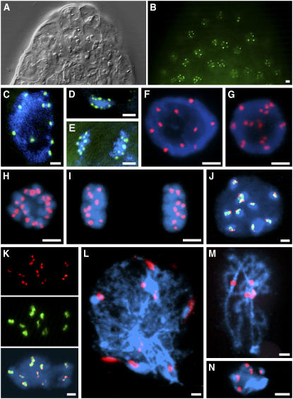
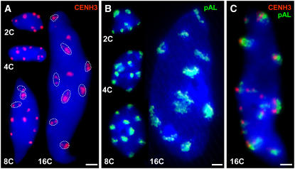
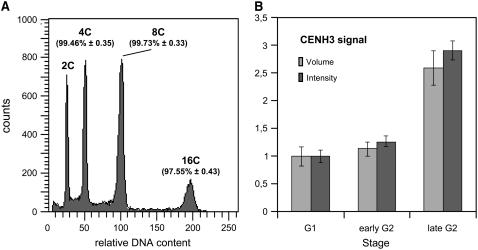
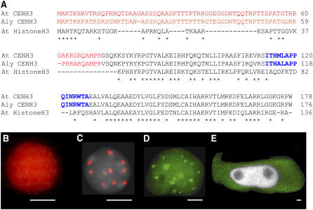
The centromeric histone H3 (CENH3) substitutes histone H3 within the nucleosomes of active centromeres in all eukaryotes. CENH3 deposition at centromeres is needed to assemble the kinetochore, a complex of conserved proteins responsible for correct chromosome segregation during nuclear division. Histones of regular nucleosomes are loaded during replication in S phase, while CENH3 deposition deviates from this pattern in yeast, human, and Drosophila melanogaster cells. Little is known about when and how CENH3 targets centromeric loci. Therefore, we determined the location and quantity of recombinant enhanced yellow fluorescent protein (EYFP)-CENH3 in mitotic root and endopolyploid leaf nuclei of transgenic Arabidopsis thaliana cells. Our data indicate significant loading of A. thaliana CENH3 during G2 (before splitting into sister kinetochores) rather than during the S or M phase of the cell cycle. The histone fold domain of the C-terminal part of CENH3 is sufficient to target A. thaliana centromeres. A. thaliana EYFP-CENH3 can recognize and target three different centromeric repeats of Arabidopsis lyrata but not field bean (Vicia faba) centromeres.


Figures

Figure 1.
Figure 1.
Localization of EYFP-CENH3 in Nuclei of Transgenic A. thaliana Plants. (A) Differential interference contrast image of an A. thaliana root tip. (B) The same root tip with EYFP signals. (C) to (E) EYFP signals in living interphase (C), metaphase (D), and anaphase (E) nuclei. (F) to (K) Immunostaining with anti-GFP antibodies (red) and/or FISH with centromeric ∼180-bp repeats (green) on meristematic root tip nuclei: early G2 (F), late G2 (G), prometaphase (H), telophase (I), early G2 showing positional coincidence of immunosignals for EYFP-CENH3, FISH signals for the ∼180-bp centromeric repeat, and bright DAPI-stained chromocentres (J), and late G2 showing colocalization of EYFP-CENH3 double dots (red) with centromeric ∼180-bp repeats (green) (K). (L) to (N) Immunostaining of EYFP-CENH3 on meiotic chromosomes: leptotene (L), pachytene (M), and diakinesis (N). DNA is counterstained with DAPI (blue). Bars = 2 μm.
Figure 2.
Figure 2.
Protein Gel Blot Analysis of Flower Buds from A. thaliana Wild-Type (Columbia) and Transgenic Lines (16, 46, and 47) Expressing the EYFP-CENH3 Fusion Protein Using Antibodies against A. thaliana CENH3. Only endogenous CENH3 yielded a band of ∼25 kD. The larger recombinant protein (∼51 kD, arrowhead) remains below the level of detection; this was also true when anti-GFP antibodies were applied.
Figure 3.
Figure 3.
Number and Appearance of EYFP-CENH3–Specific Immunosignals and Centromere-Specific FISH Signals in A. thaliana Nuclei of Different Endopolyploidy Levels. Sorted nuclei after immunofluorescence labeling of EYFP-CENH3 (A), FISH detection of centromeric ∼180-bp repeats (B), and the combination of both in a 16C nucleus (C). DNA is counterstained with DAPI (blue). In 2C to 8C nuclei, immunofluorescence and FISH signals are mainly compact, while in 16C, signals were often disperse or split into smaller foci (circled). Bars = 2 μm.
Figure 4.
Figure 4.
DNA Increase at Various Endopolyploidy Levels and Relative Proportions of CENH3 in A. thaliana Wild-Type Nuclei. (A) Histogram showing the relative DNA content (±se) of leaf nuclei after DAPI staining and flow cytometric analysis (n = 10) for distinct ploidy levels as percentages of the predicted amounts for a complete duplication of the genome per endocycle. (B) Relative proportions of CENH3 immunosignals (obtained with CENH3-rhodamine antibodies) in presumed early G2 and late G2 versus G1 nuclei (see Table 2) of A. thaliana root tip meristems expressed as signal volumes and signal intensity (±se), respectively (n = 10).
Figure 5.
Figure 5.
CENH3 sequences and A. thaliana CENH3 Localization in Nuclei of Transgenic A. thaliana, A. lyrata, and V. faba Cells. (A) Sequence alignment of At CENH3, Aly CENH3, and At Histone H3. The N-terminal parts are in red, C-terminal histone fold domain is in black, loop1 regions are in blue, and asterisks indicate sequence identity. (B) and (C) Immunolocalization of N-terminal (B) and C-terminal (C) parts of A. thaliana CENH3 fused with EYFP by anti-GFP antibodies (red) and counterstaining with DAPI (gray). (D) In vivo fluorescence (16 spots) in an A. lyrata nucleus transiently transformed with the EYFP-CENH3 construct. (E) In vivo fluorescence (green) of a Vicia faba hairy root cell transgenic for EYFP-CENH3; the nucleus (gray) remained unlabeled. Bars = 2 μm for (B) to (E).

References

    1. Ahmad, K., and Henikoff, S. (2001). Centromeres are specialized replication domains in heterochromatin. J. Cell Biol. 153 101–110. - PMC - PubMed
    1. Allshire, R.C. (1997). Centromeres, checkpoints and chromatid cohesion. Curr. Opin. Genet. Dev. 7 264–273. - PubMed
    1. Amor, D.J., and Choo, K.H. (2002). Neocentromeres: Role in human disease, evolution, and centromere study. Am. J. Hum. Genet. 71 695–714. - PMC - PubMed
    1. Amor, D.J., Kalitsis, P., Sumer, H., and Choo, K.H. (2004). Building the centromere: From foundation proteins to 3D organization. Trends Cell Biol. 14 359–368. - PubMed
    1. Badie, C., Itzhaki, J.E., Sullivan, M.J., Carpenter, A.J., and Porter, A.C. (2000). Repression of CDK1 and other genes with CDE and CHR promoter elements during DNA damage-induced G(2)/M arrest in human cells. Mol. Cell. Biol. 20 2358–2366. - PMC - PubMed

Publication types

Associated data