. 2006 Oct;18(10):2554-66.
doi: 10.1105/tpc.106.041343. Epub 2006 Oct 6.

A posttranslationally regulated protease, VheA, is involved in the liberation of juveniles from parental spheroids in Volvox carteri

Affiliations

A posttranslationally regulated protease, VheA, is involved in the liberation of juveniles from parental spheroids in Volvox carteri

Kazutake Fukada et al. Plant Cell. 2006 Oct.

Abstract

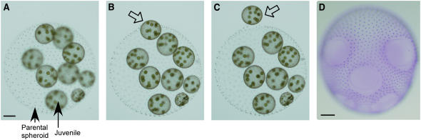
The lineage of volvocine algae includes unicellular Chlamydomonas and multicellular Volvox in addition to their colonial relatives intermediate in size and cell number. In an asexual life cycle, daughter cells of Chlamydomonas hatch from parental cell walls soon after cell division, while Volvox juveniles are released from parental spheroids after the completion of various developmental events required for the survival of multicellular juveniles. Thus, heterochronic change in the timing of hatching is considered to have played an important role in the evolution of multicellularity in volvocine algae. To study the hatching process in Volvox carteri, we purified a 125-kD Volvox hatching enzyme (VheA) from a culture medium with enzymatic activity to degrade the parental spheroids. The coding region of vheA contains a prodomain with a transmembrane segment, a subtilisin-like Ser protease domain, and a functionally unknown domain, although purified 125-kD VheA does not contain a prodomain. While 143-kD VheA with a prodomain is synthesized long before the hatching stage, 125-kD VheA is released into the culture medium during hatching due to cleavage processing at the site between the prodomain and the subtilisin-like Ser protease domain, indicating that posttranslational regulation is involved in the determination of the timing of hatching.


Figures

Figure 1.
Figure 1.
Hatching of Juveniles from Parental Spheroid. (A) to (C) Selected images taken during the hatching stage, showing a prehatched spheroid (A), liberation of a juvenile in progress ([B], arrow), and liberated juvenile leaving the parental spheroid (C). Complete animation is presented in Supplemental Movie 1 online in which images were taken every 8 s for 6.5 min and then compressed into a 19-s movie. (D) A Coomassie blue–stained parental spheroid just after liberation of all juveniles, showing openings in the parental somatic sheet. Bars = 0.1 mm.
Figure 2.
Figure 2.
VheA Purification. Pooled active fractions from DEAE Toyopearl 650M anion exchange chromatography (A) and Source 15Q anion exchange chromatography (B) and successive fractions from Superdex 200HR 10/30 gel filtration chromatography (C) were separated in a 7.5% SDS-polyacrylamide gel ([A] and [B]) or in a 4 to 20% gradient SDS-polyacrylamide gel (C) and then gels were silver stained. Numbers above the gel indicate fraction numbers, and numbers below represent ratio of released juveniles to total in the VheA assay.
Figure 3.
Figure 3.
Strategies for Cloning vheA cDNA. Arrowheads mark locations and directions of primers. The coding region and UTRs are indicated by a closed box and lines, respectively. Thick lines indicate probes used for RNA gel blot (Northern) and DNA gel blot (Southern) analyses.
Figure 4.
Figure 4.
Nucleotide Sequence and Deduced Amino Acid Sequence of vheA cDNA. A cleavage processing site and stop codon are shown by an arrowhead and an asterisk, respectively. Putative N-glycosylation sites and tandem repeat sequences in 3′-UTR are indicated by open and shaded boxes, respectively. The putative polyadenylation signal UGUAA is underlined.
Figure 5.
Figure 5.
VheA Is a Type II Membrane Protein and Subtilisin-Like Ser Protease. (A) Hydropathy plot of deduced amino acid sequence of VheA. The algorithm of Kyte and Doolittle (1982) was used with averaging over a window of nine residues. The hydrophobic region is shown by a bar. (B) Purified 125-kD VheA was separated by SDS-PAGE and analyzed by the modified PAS method. The 125-kD VheA was detected as a glycoprotein under UV illumination (left) and then stained by Coomassie blue (right). (C) Comparisons of the deduced amino acid sequence of the catalytic portion of VheA with Tg SUB1, thermitase, subtilisin Carlsberg, and hypothetical protein in C. reinhardtii genome scaffold_1. Protein sequences were aligned with a T-COFFEE algorithm (Notredame et al., 2000). Identically conserved amino acid residues, highly conserved amino acid residue, and weakly conserved amino acid residues are indicated by asterisks, colons, and periods, respectively. The catalytic triad of His, Asp, and Ser is shaded. (D) Amino acid sequence alignment of calcium binding sites of thermitase with VheA using the Blosum62 algorithm (Henikoff and Henikoff, 1992). Strong and medium-strength calcium binding sites of thermitase are shown by filled and shaded boxes, respectively. (E) Domain structure of VheA. Relative position of transmembrane segment, prodomain, subtilisin-like Ser protease domain, functionally unknown domain, and putative N-glycosylation sites (represented by Y) are indicated.
Figure 6.
Figure 6.
vheA Is a Single-Copy Gene. Genomic DNA of V. carteri was digested with HindIII (H), PstI (P), or BglII (B) and hybridized with the vheA probe shown in Figure 3.
Figure 7.
Figure 7.
vheA mRNA Begins to Accumulate Long before Hatching Stage. (A) Accumulation pattern of vheA mRNA detected by RNA gel blot hybridization using poly(A)+ RNA from various developmental stages. The probe used is shown in Figure 3. Probing for ribosomal protein S18 mRNA was performed as a loading control. (B) Quantified signal intensities on blots were normalized using the signal of S18 mRNA as a loading control. Normalized values relative to that of the 36-h time point were plotted as a function of time during the asexual life cycle. Values are means ± sd of results from three independent experiments.
Figure 8.
Figure 8.
Expression of vheA Is Specific to Juveniles. (A) Semiquantitative RT-PCR analysis of vheA and S18 expression in parental somatic cells (P) and juveniles (J). Numbers above gels indicate PCR cycles. Minus-RT controls showed no genomic DNA contamination in original RNA samples. (B) Semiquantitative RT-PCR analysis of vheA and S18 expression in juvenile somatic cells (S) and juvenile gonidia (G). (C) Schematic representation of cell types in a prehatched spheroid.
Figure 9.
Figure 9.
143-kD VheA with Transmembrane Segment Is Synthesized Long before Hatching Stage and Then 125-kD VheA Is Released into Culture Medium Due to Cleavage Processing. (A) Immunoblot analysis of cellular extracts before hatching and culture medium after hatching with anti-VheA antibody. (B) Immunoblot analysis of cellular extracts at various developmental stages. Numbers above blot indicate time points when samples were prepared. Cellular extracts before hatching (0 to 35 h) were prepared from intact spheroids, although those after hatching (36 to 42 h) were prepared from a mixture of hatched juveniles and parental somatic cells. As an internal control, actin was detected (bottom panel). (C) Immunoblot analysis of culture medium samples collected at various developmental stages.

References

    1. Aono, N., Inoue, T., and Shiraishi, H. (2005). Genes specifically expressed in sexually differentiated female spheroids of Volvox carteri. J. Biochem. (Tokyo) 138 375–382. - PubMed
    1. Araki, K., Fujikawa, N., Nakayama, I., Nagoya, H., and Onozato, H. (1996). Early expression of a hatching enzyme gene in masu salmon (Oncorhynchus masou) embryos. Can. J. Fish. Aquat. Sci. 53 509–512.
    1. Bendtsen, J.D., Nielsen, H., von Heijne, G., and Brunak, S. (2004). Improved prediction of signal peptides: SignalP 3.0. J. Mol. Biol. 340 783–795. - PubMed
    1. Burge, C., and Karlin, S. (1997). Prediction of complete gene structures in human genomic DNA. J. Mol. Biol. 268 78–94. - PubMed
    1. Cheng, Q., Pappas, V., Hallmann, A., and Miller, S.M. (2005). Hsp70A and GlsA interact as partner chaperones to regulate asymmetric division in Volvox. Dev. Biol. 286 537–548. - PubMed

Publication types

MeSH terms